a belt of extrusomes running meridionally from oral bulge
belt of extrusomes running from oral bulge over ventral side to the mid of dorsal side
symbiotic algae present, often filled with dark granules
contactile vacuole subterminal
macronucleus oval, elongate or ribbon-like with an adjacent micronucleus
soft, long cilia, slowly swimming
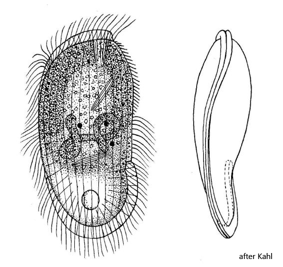
Penardiella interrupta
I found this haptorian ciliate in the uppermost (still aerobic) mud layer in the Simmelried.(2009) and in June 2025 again the pond behind parking space St. Ulrich (Austria). Penardiella interrupta is somewhat difficult to identify because a thoroughly investigation of the ciliate is necessary to make the assignment certain. Like Spathidium, Penardiella has an oral bulge armed with extrusomes. From the oral bulge, a broad, double-row belt of extrusomes runs over the ventral side, encircles the posterior end und reaches up to the middle of the dorsal side, where the belt ends (s. figs. 5 a-b, 6, 7 a-b and 8). Penardiella interrupta can be reliably identified by this characteristic.
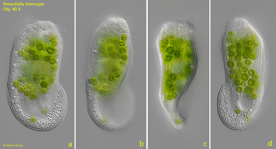
Fig. 1 a-c: Penardiella interrupta. L = 118 µm. A freely swimming specimen. Obj. 40 X.
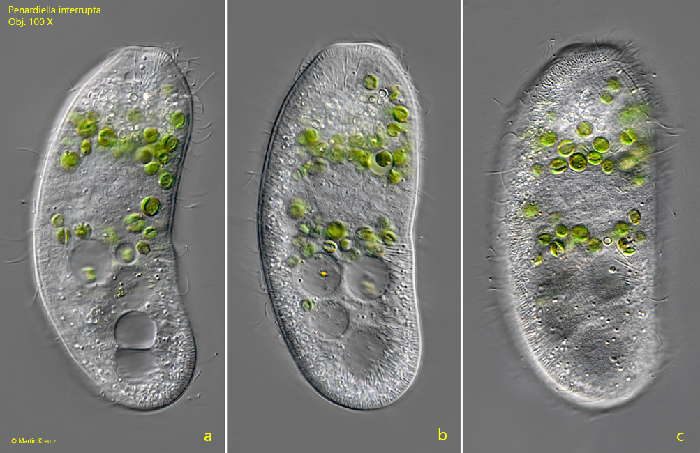
Fig. 2 a-c: Penardiella interrupta. L = 100 µm. A second freely swimming specimen in lateral view from left. Obj. 100 X.
Fig. 3: Penardiella interrupta. L = 100 µm. A second freely swimming specimen in lateral view from left. Obj. 100 X.
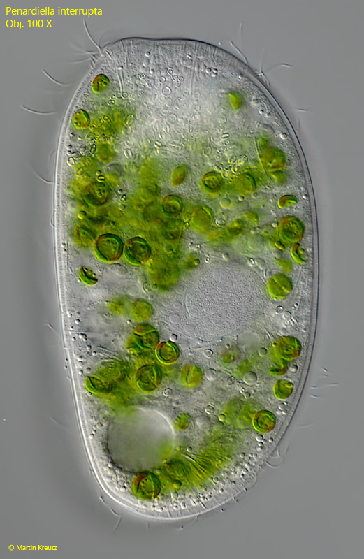
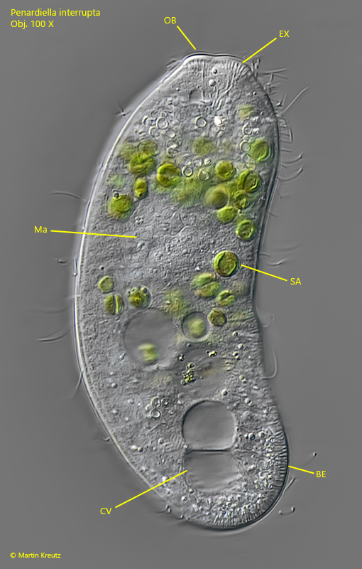
Fig. 4: Penardiella interrupta. L = 100 µm. A freely swimming specimen in lateral view from left. BE = belt of extrusomes, CV = contractile vacuole, EX = extrusomes, Ma = macronucleus, OB = oral bulge. Obj. 100 X.
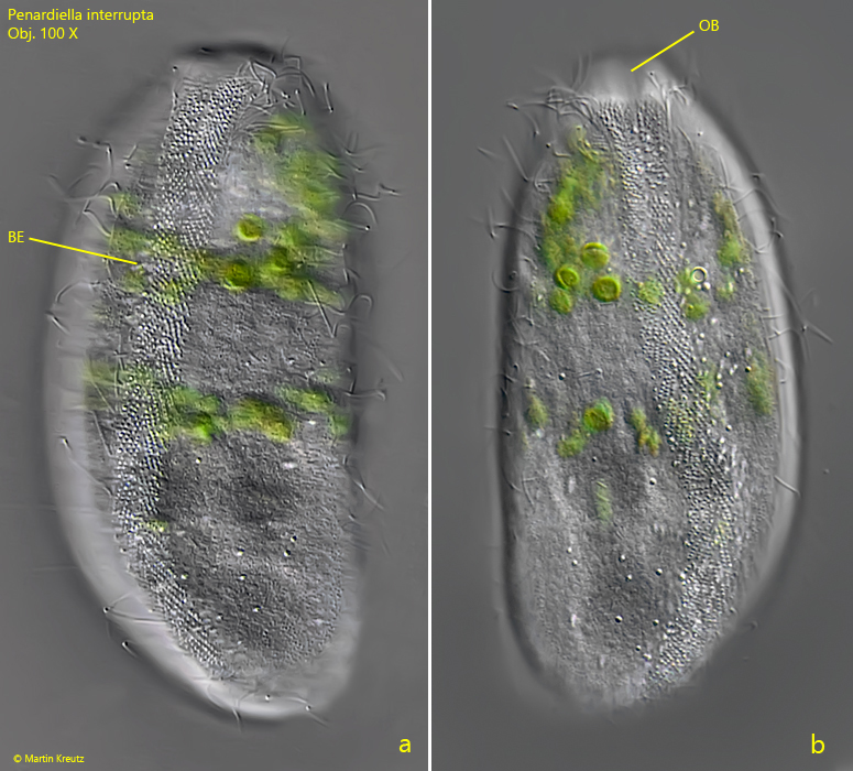
Fig. 5 a-b: Penardiella interrupta. L = 100 µm. A slightly squashed specimen in ventral view. From the oral bulge a belt of extrusomes (about 10 µm wide) runs meridionally across the ventral side and encircles the posterior end. BE = belt of extrusomes, OB = oral bulge. Obj. 100 X.
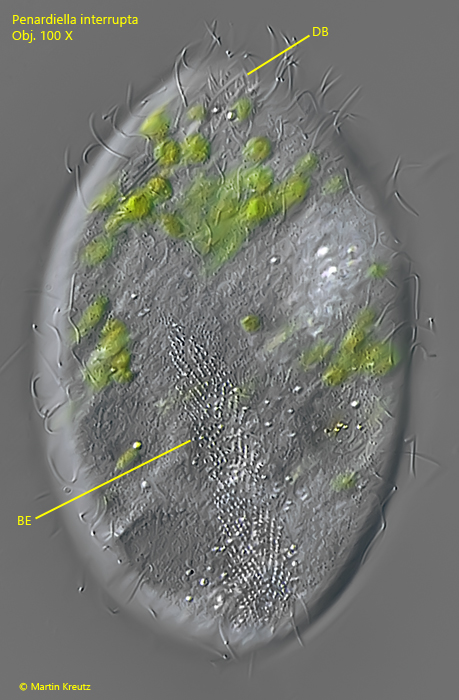
Fig. 6: Penardiella interrupta. L = 100 µm. Coming from the ventral side, the belt of extrusomes runs to the middle of the dorsal side and ends there. BE = belt of extrusomes, DB = dorsal brush. Obj. 100 X.
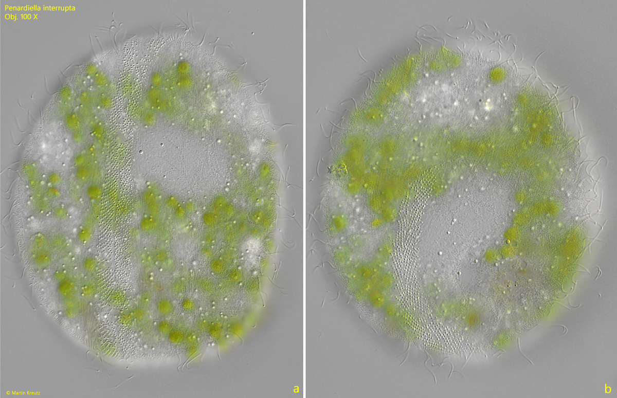
Fig. 7 a-b: Penardiella interrupta. A squashed specimen from ventral (a) and dorsal (b). Obj. 100 X.
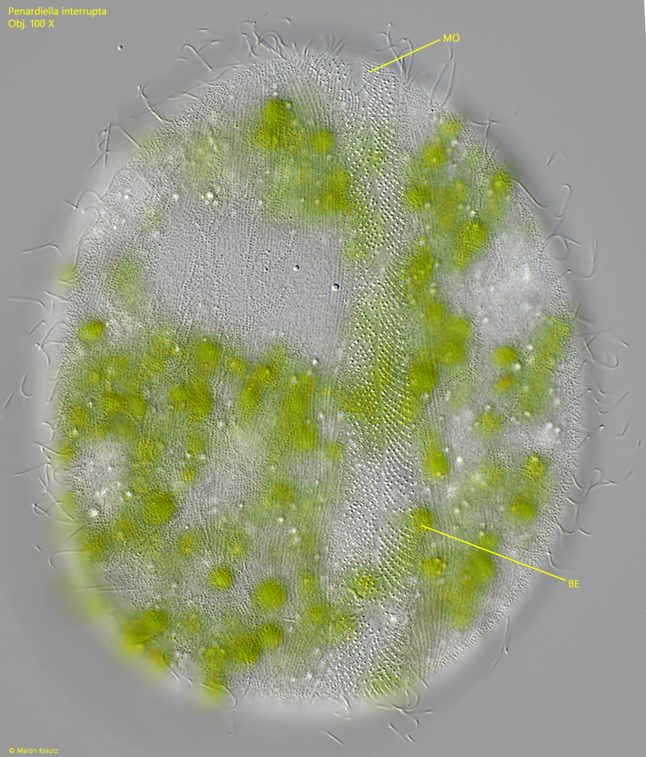
Fig. 8: Penardiella interrupta. The ventral belt of extrusomes (BE) in detail. MO = mouth opening. Obj. 100 X.
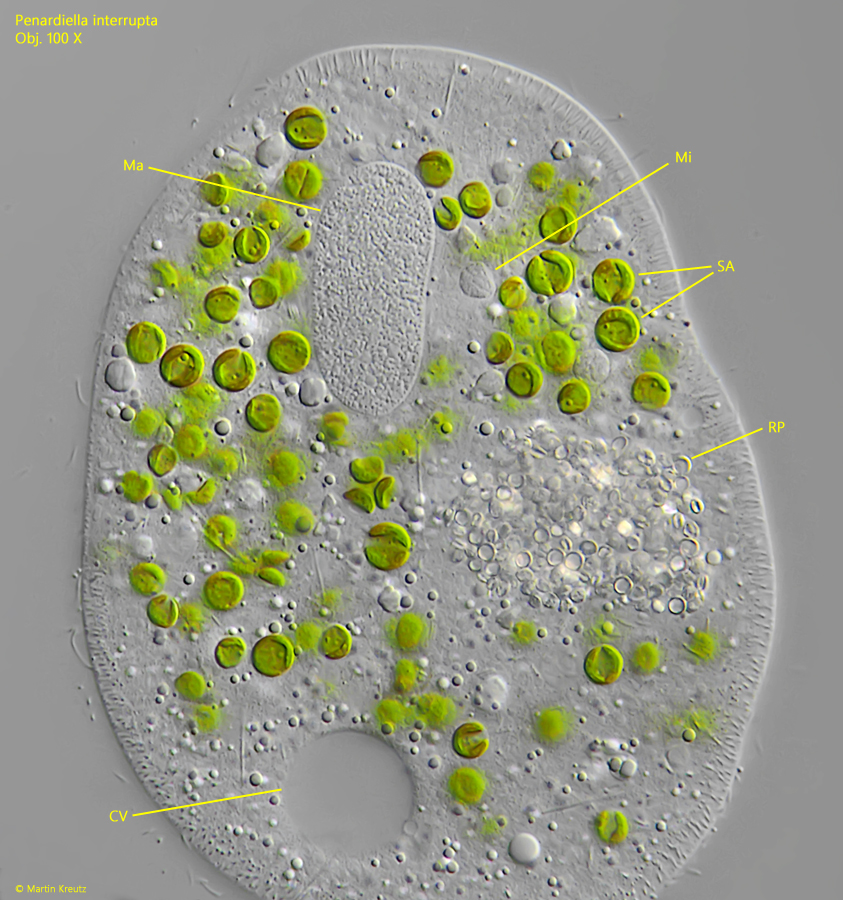
Fig. 9: Penardiella interrupta. L = 100 µm. The macronucleus (Ma) and micronucleus (Mi) in a strongly squashed specimen. Near mid body the remains of prey are visible (RP), likely the ring-shaped granules of Dexiotricha granulosa. CV = contractile vacuole, SA = symbiotic algae. Obj. 100 X.
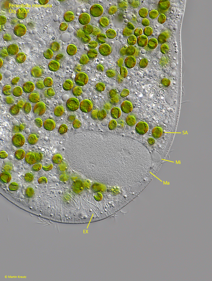
Fig. 10: Penardiella interrupta. The macronucleus (Ma) and micronucleus (Mi) in a second specimen. EX = extrusomes, SA = symbiotic algae. Obj. 100 X.