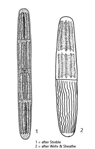
cells cylindrically, attenuating gradualley to truncate or rounded apices
length 80–400 µm
cell wall with spiralized ridges
2–4 chlorplasts per cell
chloroplasts with 6–7 longitudinal ridges and 1–3 pyrenoids
pyrenoids somtimes elongated
girdle bands present
nucleus central
terminal vacuoles in the apices absent
Penium spirostriolatum
I found Penium spirostriolatum in June 1995 in the Ibmer Moor (Austria) and in 2025 in the Schwemm Moor (Austria). The alga is not present in my local sites.
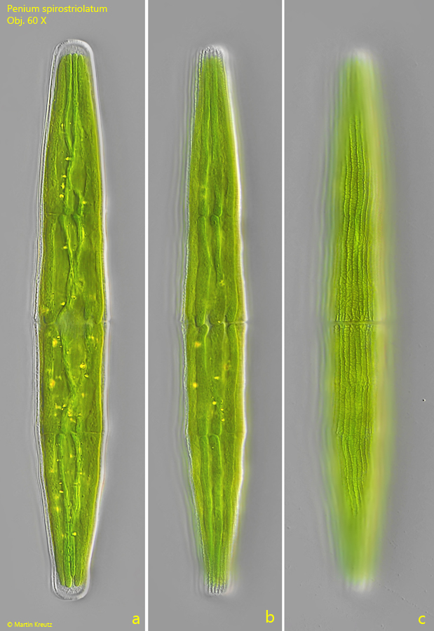
Penium spirostriolatum can easily be recognized by the 4 chloroplasts per cell, which are separated from each other by transverse gaps (s. fig. 1 b). The chloroplasts have 6–7 longitudinal ridges, making them appear star-shaped in cross-section. The pyrenoids in the chloroplasts can sometimes have an elongated, stretched shape.
The similar species Closterium closteroides var. intermedium can also have 4 chloroplasts, which are separated by transverse gaps. However, this species has clear teminal vacuoles at the apices, in which crystals are visible, as is typical for the genus Closterium.
Fig. 1 a-b:Penium spirostriolatum. L = 210 µm. Three focal planes of a specimen from the Schwemm Moor (Austria). Obj. 60 X.
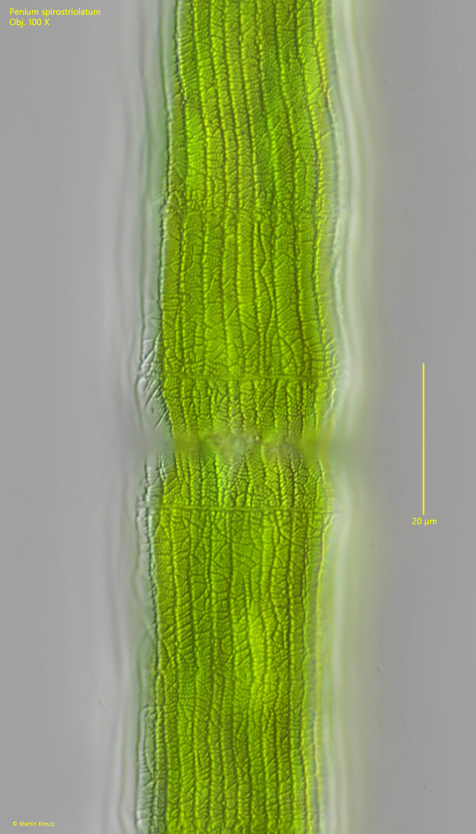
Fig. 2:Penium spirostriolatum. The network structure of the cell wall in detail. Obj. 60 X.