ridge runs at first to left side, over dorsal side to right side, turn back to ventral side and reach the right side again at posterior end
body often filled with oily droplets and granules
contractile vacuole terminal
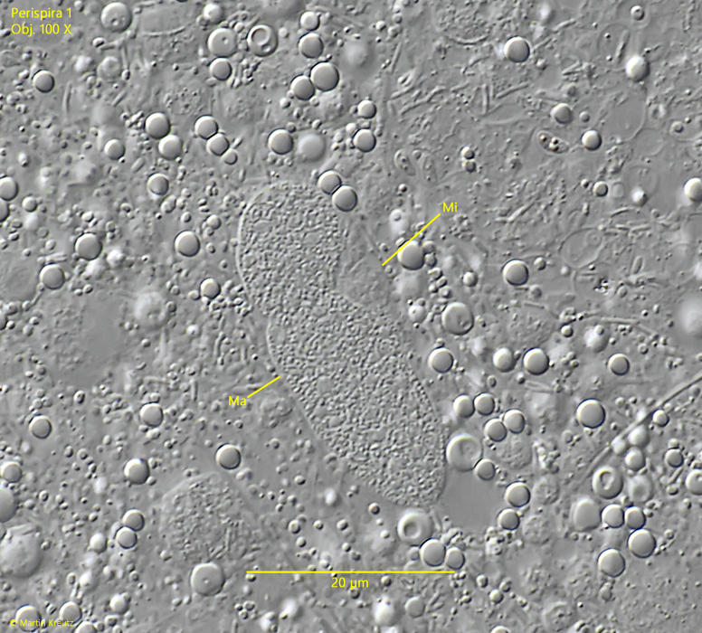
macronucleus ellipsoid
one small micronucleus
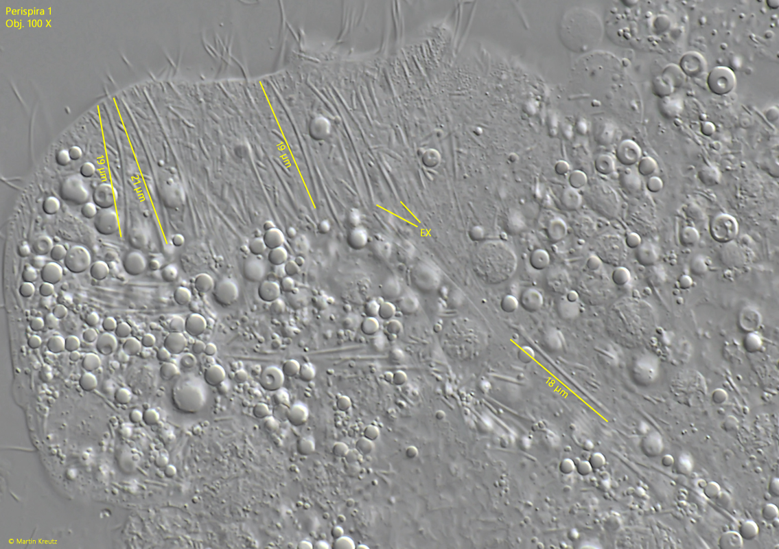
extrusomes slightly curved rods, 18–21 µm long
No drawings from previous authors vailable.
Between 2003 and 2012, I found 6 specimens of a ciliate that possesses the characteristics of the genus Perispira. I found all specimens during the summer months from June to August in the years 2002–2022. Five specimens came from the upper mud layer in the Simmelried and one specimen (2014) from the Ulmisried.
The essential characteristic of the genus Perispira is a clearly visible ridge of extrusomes, which runs in a spiral line from the anterior to the posterior end of the body. This principal course of the ridge is the same in the three species described so far: Perispira ovum, Perispira strephosoma, Perispira carinata and Perispira pyriformis.
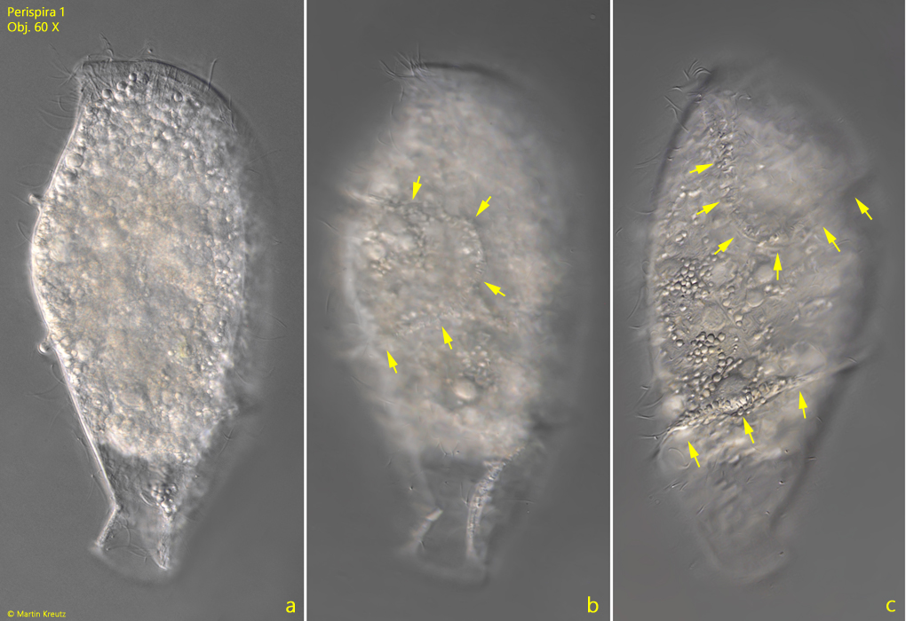
In the specimens I examined, however, the course of the ridge fundamentally differs from that of the previously described species. It begins at the anterior end and already bends to the right side in the anterior quarter (s. fig. 1 c). There, the ridge describes a clearly visible 180° curve (s. figs. 1 b and 2 b) and returns below the equator to the ventral side to then encircle it (s. figs. 1 c and 2 c) and end on the dorsal side at the posterior end. This rather complex course differs so significantly from the previously described species that it must be a species not yet described. I provisionally designate it as Perispira 1. In addition to the unusual course of the ridge, the specimens of Perispira 1 are larger, measuring 130–180 µm, compared to the previously described species, which range between 70–130 µm.
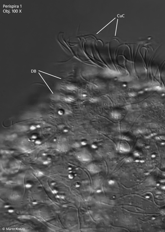
The macronucleus of Perispira 1 is short ellipsoid with a small, attached micronucleus (s. fig. 6). The extrusomes of Perispira 1 are slightly curved, thin rods with a length of 18–21 µm (s. fig. 7). The dorsal brush is three-rowed and very short (s. fig. 8). The apical ridge of extrusomes appears to be surrounded by curved cilia (s. fig. 8), as Penard and Kahl described for Perispira strephosoma. However, this was only observed apically and not along the further course of the ridge as in Perispira strephosoma.
Fig. 1 a-c:Perispira 1. L = 180 µm. A second freely swimming specimen from right (a, b) and from ventral (c). The specimen was found in August 2008 in the Simmelried. The ridge of extrusomes starts anteriorly and run at first to the right side (c, upper arrows) were it describe a 180° curve (b, arrows) and crosses the ventral side in the posterior half (c, lower arrows) before it reach the posterior end on the dorsal side. Obj. 60 X.
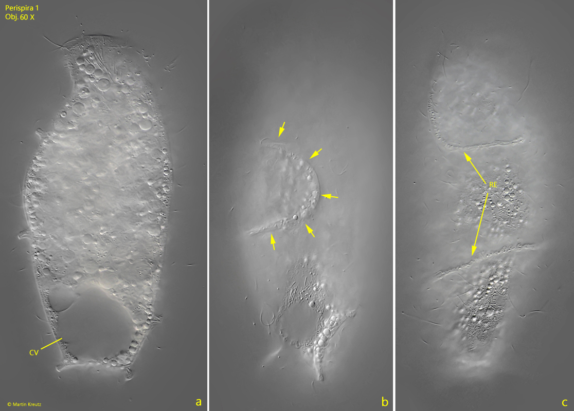
Fig. 2 a-c:Perispira 1. L = 145 µm. A second freely swimming specimen from right (a, b) and from front right (c) found in July 2022 in the Simmelried. Note the 180° curve of the ridge of extrusomes (RE) on the right side (b). CV = contractile vacuole. Obj. 60 X.
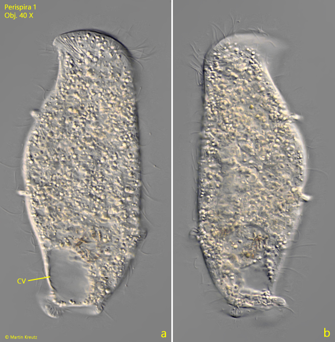
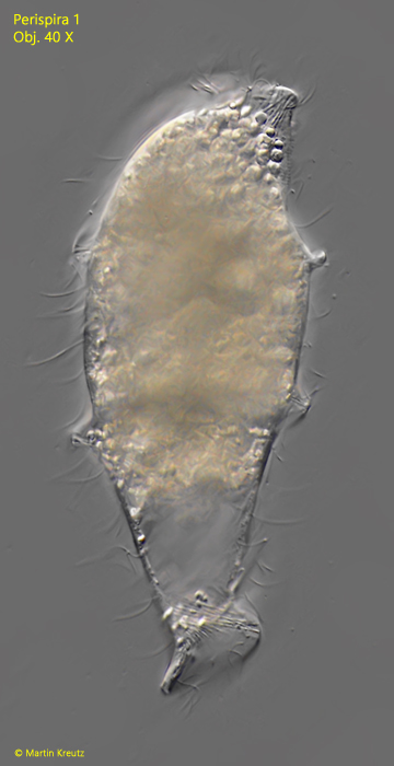
Fig. 3 a-b:Perispira 1. L = 171 µm. A third freely swimming specimen from right (a) and from left (b). Obj. 40 X.
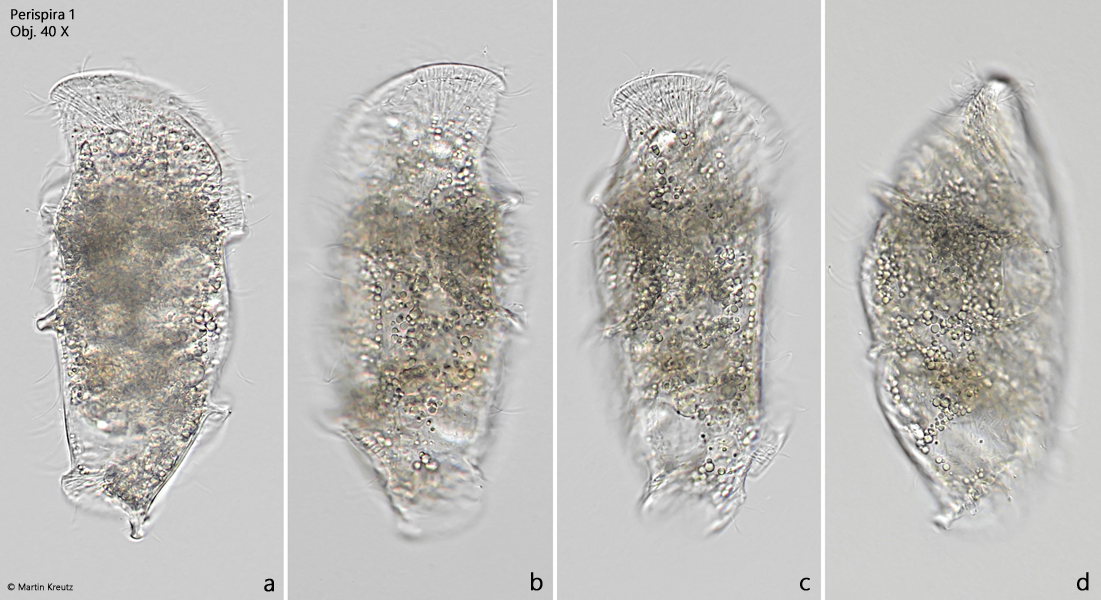
Fig. 4 a-d:Perispira 1. L = 155 µm. A freely swimming specimen in brightfield illumination from right (a, c), left (b) . ventral side in the posterior half (c, lower arrows) before it reach the posterior end on the dorsal side. Obj. 40 X.
Fig. 5:Perispira 1. L = 180 µm. The same specimen as shown in fig. 2-a-c from dorsal. Obj. 40 X.
Fig. 6:Perispira 1. The macronucleus (Ma) and the micronucleus (Mi) in a squashed specimen. Obj. 100 X.
Fig. 7:Perispira 1. The slightly curved, rod-shaped extrusomes with a length of 18–21 µm in a strongly squashed specimen. Obj. 40 X.
Fig. 8:Perispira 1. The dorsal brush (DB) in a slightly squashed specimen. The apical ridge of extrusomes is surrounded by curved cilia (CuC). Obj. 100 X.