ridge of extrusome describe a whole spirally turn over the body
dorsal inconspicuous, three rows
sometimes with symbiotic algae
nucleus horseshoe-shaped or ovoid
one spherical micronucleus
contractile vacuole terminal
feeding on Euglena species
Perispira ovum
I rarely find Perispira ovum and mostly only in the top layer of mud. The species is most commonly found in the Simmelried. So far, I have only found one specimen in the Bussenried.
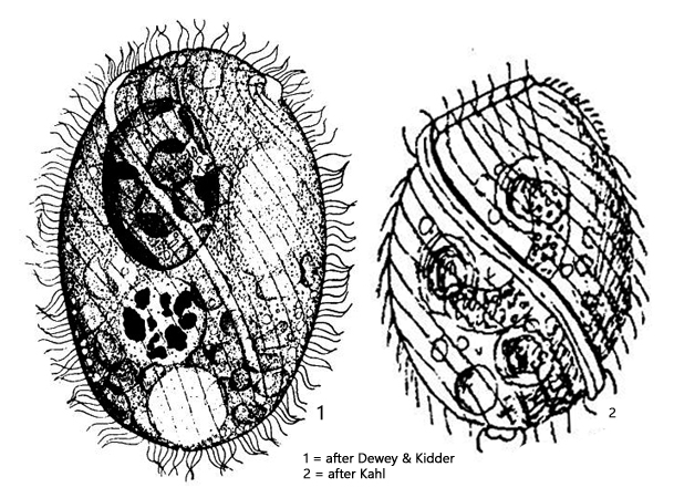
The most striking and essential feature of Perispira ovum is the spirally running ridge of the extrusomes, which begins apically and ends at the rear after a complete turn around the body (s. figs 1 a-c and 2 a-c). The specimens of my population were comparatively slender and between 60–80 µm in size. As Kahl reports, the shape strongly depends on the nutritional state. He also reports that Perispira ovum primarily feeds on Euglena viridis. Later, Johnson et al. (1995) reported that the ciliate feeds on Euglenaformis proxima (syn. Euglena proxima), and they also describe the type of prey capture of Perispira ovum. According to this, the key stimulus is probably the contact of the ridge of the extrusomes with the flagellum of Euglenaformis proxima, whereupon the flagellum sticks to the ridge and a mouth opening forms to engulf the prey. The specimens observed by Johnson et al. were all intensely green and darkly colored due to the phagocytosed euglenids. However, the specimens of my population were always colorless with some oil-like inclusions and crystals. Unfortunately, I was never able to observe the feeding process myself.
As another result of the investigations on Perispira ovum, Johnson et al. described that the ciliate incorporates the chloroplasts, mitochondria, and also the paramylon granules of the prey organisms and surrounds them with a membrane of the endoplasmic reticulum. However, it remains unclear whether these incorporated organelles continue to maintain their function in Perispira ovum.
Perispira ovum is very similar to the species Perispira strephosoma in both shape and size, but Perispira strephosoma always possesses several small micronuclei. I could only ever identify one micronucleus (s. fig. 1 e), which is why it must be Perispira ovum.
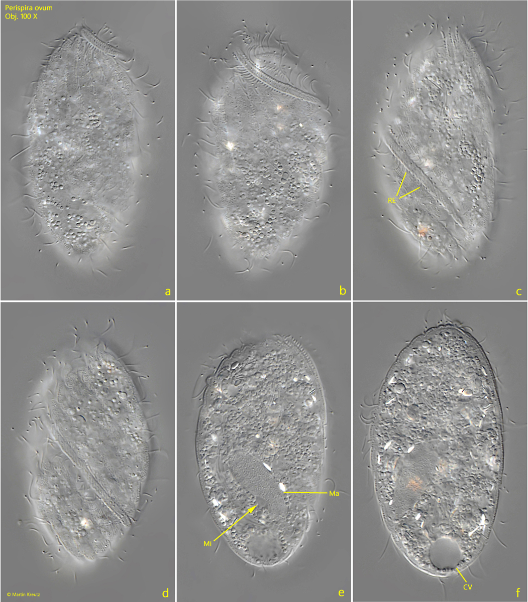
Fig. 1 a-f:Perispira ovum. L = 58 µm. A slightly squashed specimen from ventral (a), fronto-dorsal (b) and dorsal (c–f). The ridge of extrusomes (RE) describe a spirally turn around the body from the anterior to the posterior end. CV = contractile vacuole, Ma = macronucleus, Mi = micronucleus. Obj. 100 X.
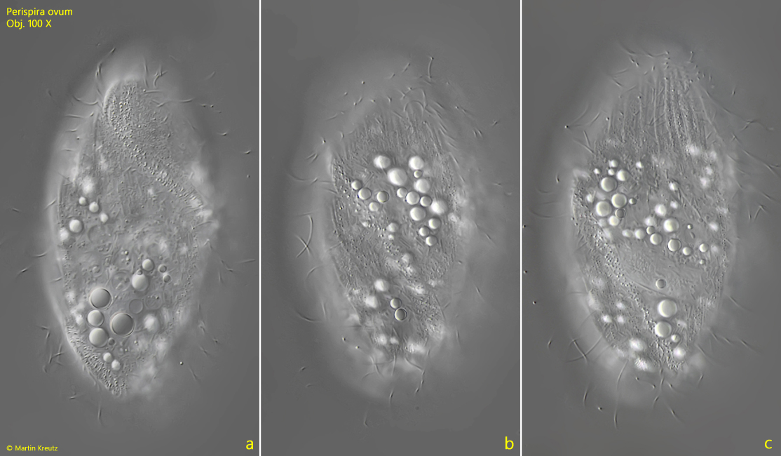
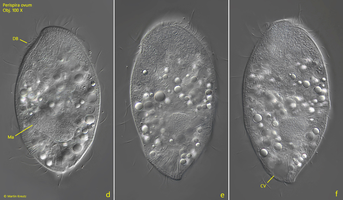
Fig. 2 a-f:Perispira ovum. L = 80 µm. A second, freely swimming specimen found in October 2018 in the Bussenried. CV = contractile vacuole, DB = dorsal brush, Ma = macronucleus. Obj. 100 X.