So far, I have only found 2 specimens of Petalomonas mira var. truncata. The first in March 1995 and the second in June 2024. Both specimens come from the Simmelried. Since then, I have not recorded any further findings.
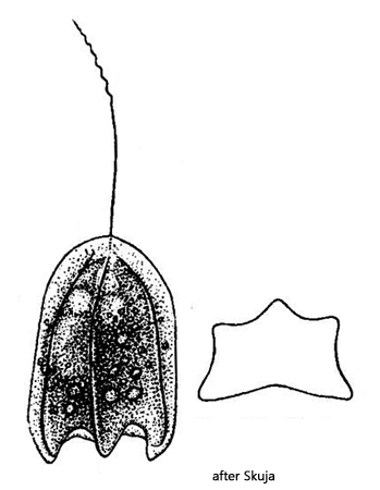
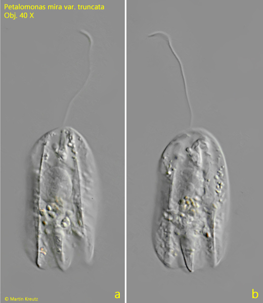
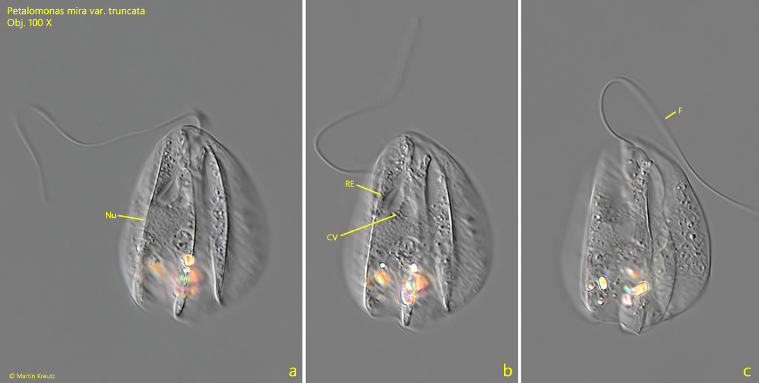
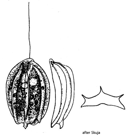
The specimen found in March 1995 corresponds in its shape in every case to the original description by Skuja (1956), especially regarding the 3 keels and the parallel lateral sides (s. fig. 1 a-b). However, the specimen was 56 µm long and thus over 50% longer than stated by Skuja. The second specimen from June 2014 was also longer at 42 µm but had a significantly broader shape with convex lateral sides (s. fig. 3 a-c). The only alternative species to consider is Petalomonas tricarinata. This species was also first described by Skuja (1939) and has a dorsal spine at the posterior end, which I could not observe in my specimens (s. fig. 4). Despite the deviations in shape and length, I therefore see no alternative to Petalomonas mira var. truncata.
Fig. 1 a-b:Petalomonas mira var. truncata. L = 56 µm. Two focal planes of a specimen found in March 1995 in the Simmelried. Obj. 40 X.
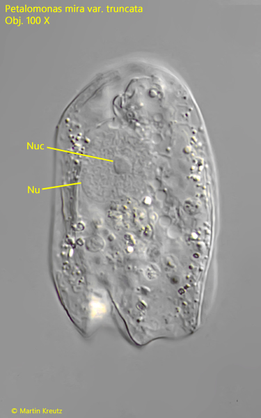
Fig. 2:Petalomonas mira var. truncata. L = 56 µm. The squashed specimen as shown in fig. 1 a-b. The nucleus (Nu) has a central nucleolus (Nuc). Obj. 100 X.
Fig. 3 a-c:Petalomonas mira var. truncata. L = 42 µm. Different focal planes from dorsal of a specimen found in June 2014 in the Simmelried. CV = contractile vacuole, F = flagellum, Nu = nucleus, RE = reservoir. Obj. 100 X.
Fig. 4:Petalomonas tricarinata. Drawing of the species by Skuja. Note the posterior spine.