Phacus alatus G.A. Klebs, 1883

Most likely ID: n.a.


Synonym: n.a.


Sampling location: Simmelried


Phylogenetic tree: Phacus alatus


Diagnosis: 

  • cell broadly ovoid, flattened
  • cell shape in two unequal halves
  • length 24–45 µm, width 19–34 µm
  • 2 prominent paramylon bodies, laterally located
  • many chloroplasts, disc-shaped
  • short and curved caudal spine
  • one large eyespot
  • one flagellum, about body length
  • striation of the pellicle longitudinally
Phacus-alatus
Phacus alatus
In April 2026 I found Phacus alatus in large numbers between floating plant material in the Simmelried. Otherwise I find this species only very rarely.

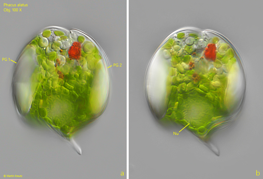
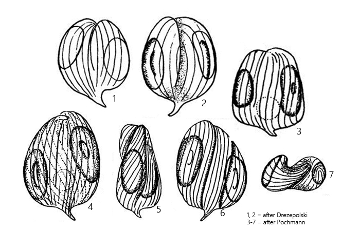
Phacus alatus has a compact shape with a sharply curved, short caudal spine. The cells often have an asymmetrical form because the two halves of the cell can be slightly different in size. Striking are two large paramylon grains, which lie laterally (s. fig. 1 a). They are so large that they deform the cell and noticeably thicken it. There are additional, much smaller paramylon grains, all of which are round. The eyespot is conspicuously large. I was not able to document the flagellum, because the specimens reacted very sensitively to the coverslip being placed on them and immediately shed the flagellum.

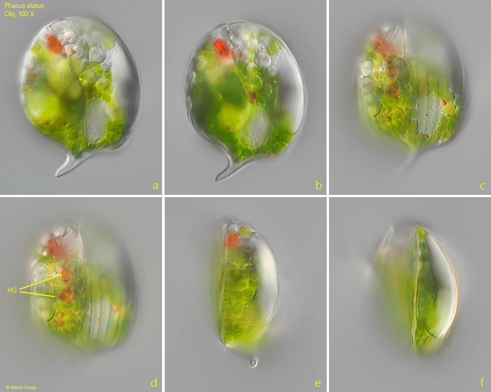
The specimens of my population were around 50 µm long. It is difficult to say what the maximum dimensions of this species are, since Phacus alatus has meanwhile been synonymized with several other (larger) species such as Phacus platyaulax or Phacus macrostigma. In the cytoplasm of the specimens of my population there were quite a lot of haematochrome granules, which were not mentioned by earlier authors (s. fig. 2 d).
Phacus-alatus

Fig. 1 a-b: Phacus alatus. L = 49 µm. A freely swimming specimen from ventral. Note the large paramylon grains (PG 1, PG 2) located laterally in the cell. Nu = nucleus. Obj. 100 X.

Phacus-alatus

Fig. 2 a-f: Phacus alatus. L = 53 µm. A second freely swimming specimen from dorsal (a-d) and from left (e, f). In the cytoplasm several orange haematochrome grains (HG) are visible. Obj. 100 X.

Phacus-alatus

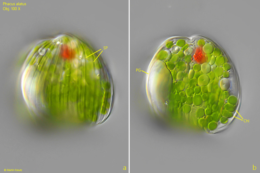
Fig. 3 a-b: Phacus alatus. A third specimen from ventral with focal planes on the longitudinal striation of the pellicle (SP) and on the disc-shaped chloroplasts (Chl). Obj. 100 X.

Phacus-alatus

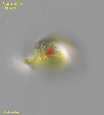
Fig. 4: Phacus alatus. Apical view on a freely swimming specimen. The shape is slightly twisted. Obj. 40 X.