cell ovoid or trapezoid, dorso-ventrally flattened
dorsal keel reach to mid-body
length 37–80 µm, width 30–50 µm
one larger and one smaller paramylon grain
chloroplasts disc-shaped
caudal spine sharply set off, oblique, straight or slightly curved
one flagellum, about body length
pellicle longitudinally striated
eyespot conspicuous
Phacus pleuronectes
I find Phacus pleuronectes frequently and regularly at some of my localities. In my population, the specimens were mostly between 50 and 80 µm in size, although specimens up to 100 µm have also been described in the literature. So the size seems to be quite variable.
I consider the dorsal keel, which extends to the middle of the body (s. figs. 3 c and 4 c), and the short, quite sharply defined caudal spine to be important identification features. It bends to the right (seen ventrally, the flat side) with an angle of about 45° in relation to the longitudinal axis of the body.
Differentiation from the similar species Phacus orbicularis is difficult. This species is almost as wide as it is long and is always said to be over 50 µm long. However, as the shape and length of Phacus pleuronectes is also variable, there may be overlaps. Kusel-Fetzmann (2002) discusses whether only specimens under 40 µm in length should be counted as Phacus pleuronectes. She also cites the delicate transverse stripes between the longitudinal stripes of the pellicle as a distinguishing feature. The specimens with transverse stripes should be classified as Phacus orbicularis and those without as Phacus pleuronectes. In my experience, however, this distinguishing feature is also variable. In my population of Phacus pleuronectes, this cross-striping was actually quite clearly visible (s. fig. 5). The transverse striation is found in many Phacus species, but is not present in all specimens or is too weakly developed to make it visible under the light microscope. Other authors have also not cited this feature as a distinguishing characteristic. I therefore consider the specimens shown below to be Phacus pleuronectes, also because of the slightly trapezoidal body shape, which narrows slightly towards the apical end.
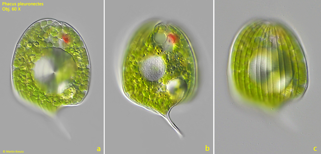
Fig. 1 a-c:Phacus pleuronectes. L = 77 µm (with spine). Different focal planes of a slightly squashed specimen from ventral. Obj. 60 X.
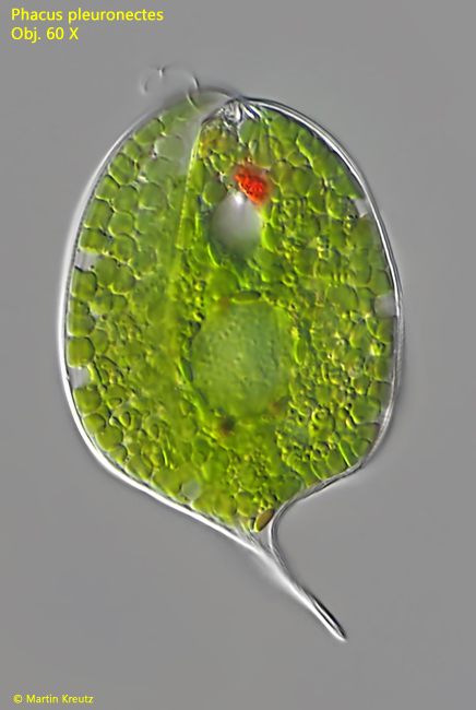
Fig. 2:Phacus pleuronectes. L = 82 µm (with spine). A second specimen from ventral. Obj. 60 X.
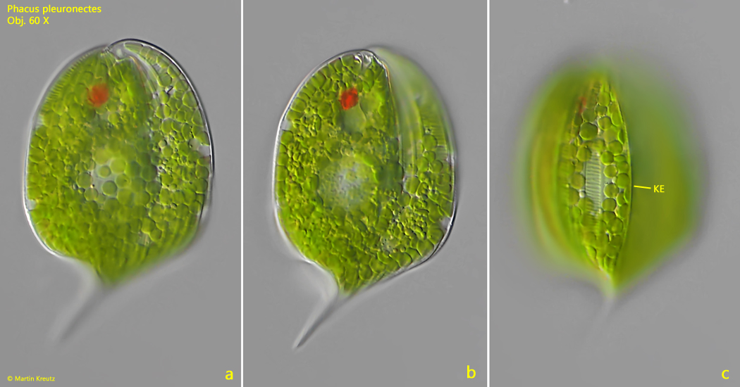
Fig. 3 a-c:Phacus pleuronectes. L = 77 µm (with spine). Different focal planes of a slightly squashed specimen from dorsal. Note the dorsal keel (KE). Obj. 60 X.
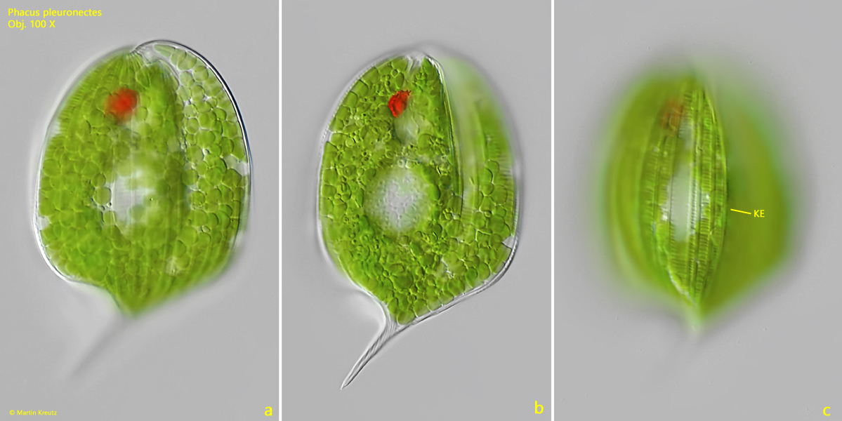
Fig. 4 a-c:Phacus pleuronectes. L = 77 µm (with spine). The same specimen as shown in fig. 3 a-c at higher magnification. KE = keel. Obj. 100 X.
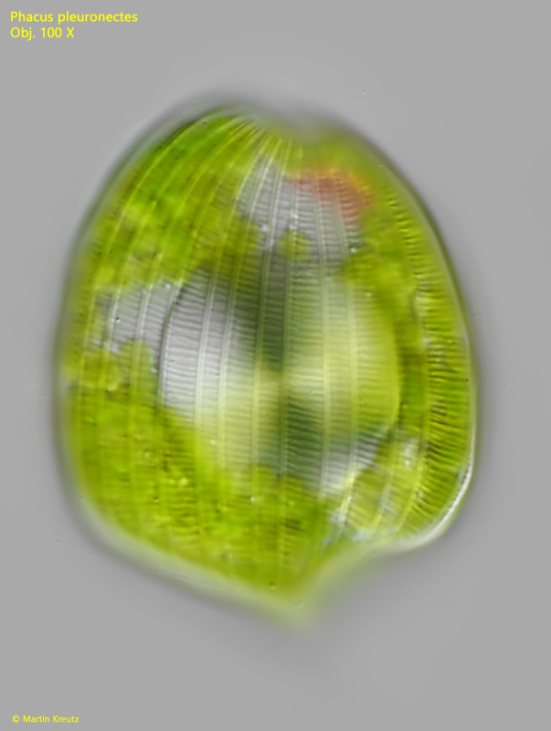
Fig. 5:Phacus pleuronectes. L = 77 µm (with spine). The flat ventral side of a specimen with focal planes on the transverse striation of the pellicle. Obj. 100 X.
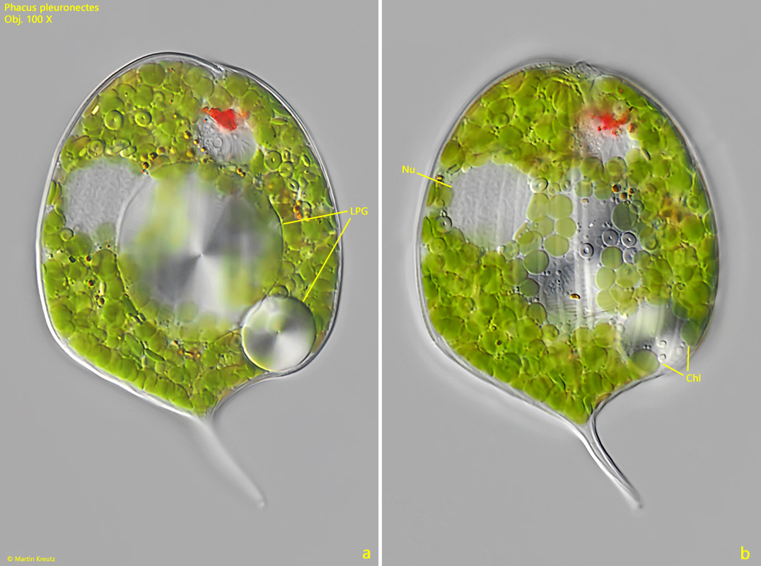
Fig. 6 a-b:Phacus pleuronectes. Two focal planes of a squashed specimen from ventral. Note the two large paramylon grains (LPG) of different size and the disc-shaped chloroplasts (Chl). Nu = nucleus. Obj. 100 X.