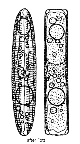
cell linear elliptical, parallel sides, rounded apices in valve view
in girdle view elongated rectangular
length 50–170 µm, width 10–30 µm
parallel longitudinal striae, asymmetric expansion in center
raphe wavy, with curved endings
two plate-shaped chloroplasts
chlorplasts golden-grown, greenish
Pinnularia viridis
Pinnularia viridis is one of the most common diatoms in my sampling areas. The specimens are found among aquatic plants, but also in the top layer of sludge on the bottom.
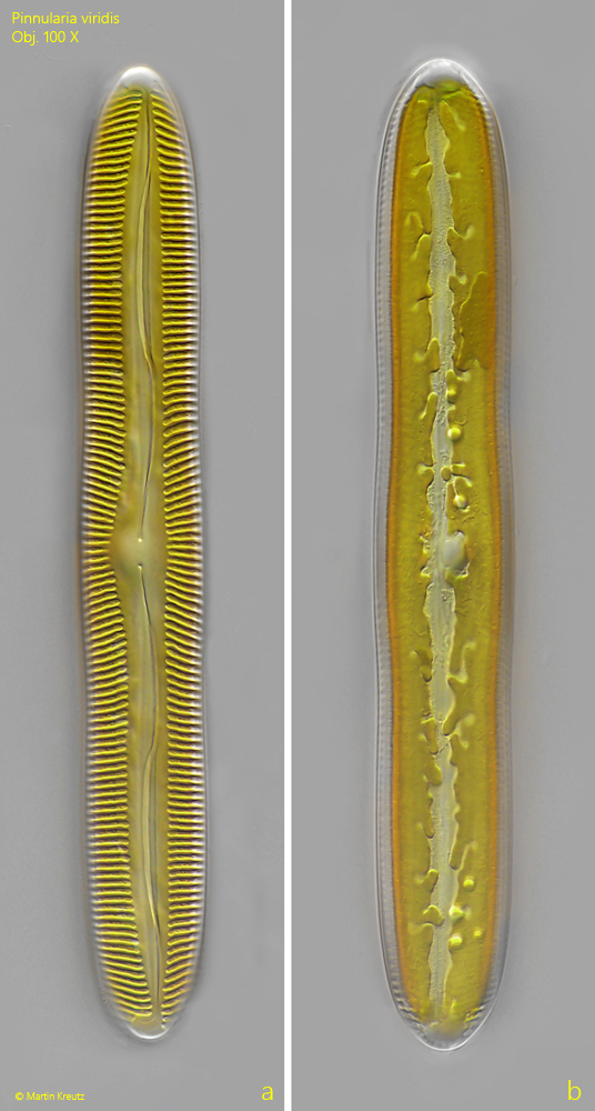
In valve view the specimens are long-elliptical with nearly parallel sides. The ends are rounded. The striation is strongly pronounced. It does not have a gap in the middle of the cell, but instead forms a somewhat asymmetrically shaped central area there (s. fig. 1 a). The raphe in the center shows a characteristic wavy shape and is bent around at the ends of the cell. The cell has two chloroplasts that lie against the girdle bands. In valve view only their lobed margins can be seen (s. fig. 1 b). The chloroplasts in my population had a yellowish-green coloration. In DIC the color changes somewhat, depending on the orientation of the specimens in the preparation.
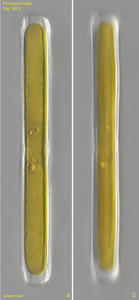
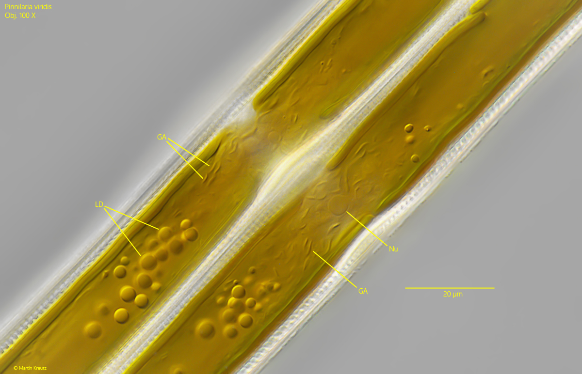
In girdle view, Pinnularia viridis is elongated-rectangular (s. fig. 2 a-b). The chloroplasts cover the entire interior surface. Oil droplets of various sizes are often present inside the cell, often also at both ends of the cell (s. fig. 3).
In Pinnularia viridis, the Golgi apparatus can be recognized under the light microscope, which is particularly distinctly developed in this diatom. In the light microscope they appear comma-shaped with a length of about 3–4 µm (s. fig. 3). That these are indeed the Golgi apparatus was shown by Schmid (1989) through comparative investigations using the light microscope and the electron microscope.
Fig. 1 a-b:Pinnularia viridis. L = 122 µm. Two focal planes of specimen in valve view. The two chloroplasts in the cell have lobed margins (b). Obj. 100 X.
Fig. 2 a-b:Pinnularia viridis. L = 150 µm. Two focal planes of a second specimen in girdle view. Obj. 100 X.
Fig. 3:Pinnularia viridis. Two specimens after cell division in girdle view. Around the cell nucleus (Nu), the comma-shaped Golgi apparatuses (GA) are arranged. LD = lipid droplets. Obj. 100 X.