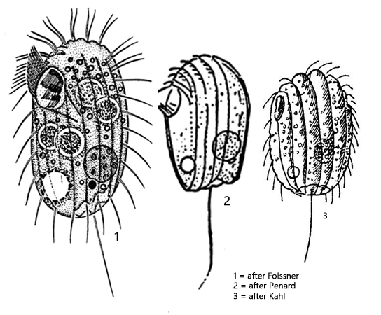
posterior end with indentation surrounded by with knobby margin
pellicle stiff with longitudinal ridges
mouth opening small, anterior third
oral apparatus with 3 adoral membranelles and a undulating membrane
14 longitudinale rows of cilia between ridges
length 30–50 µm, width 17–30 µm
macronucleus spherical, posterior half, dorsal
one micronucleus, adjacent to macronucleus
contractile vacuole posterior third, ventral side
one caudal cilium
Platynematum sociale
According to Kahl (1935), Platynematum sociale is supposed to be very common, especially in eutrophic waters. However, I find Platynematum sociale only very rarely and always only single specimens. Foissner et al. (1994) were also only able to find the species once in the iron oxide sludge of a small pond.
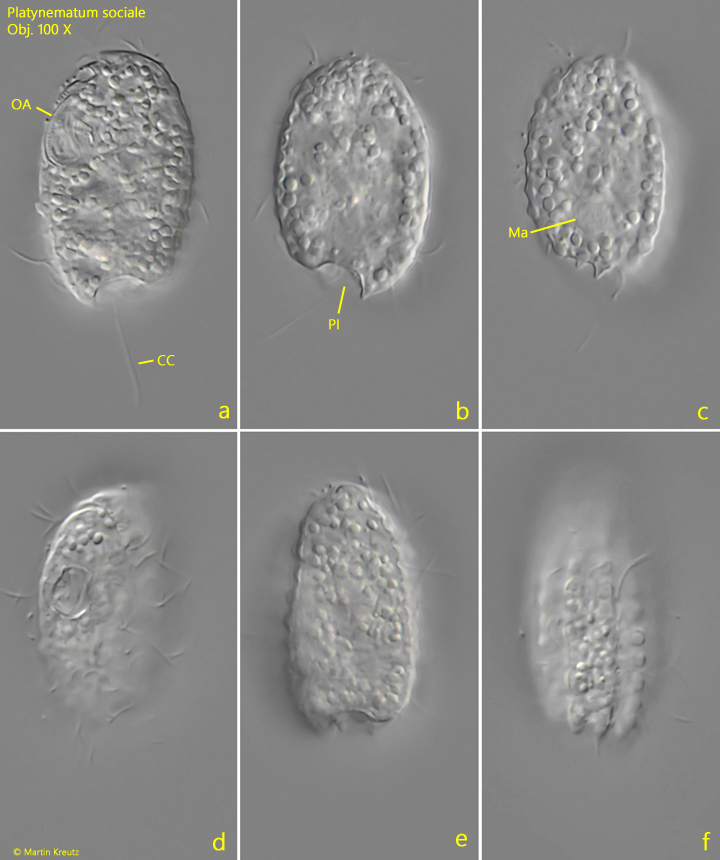
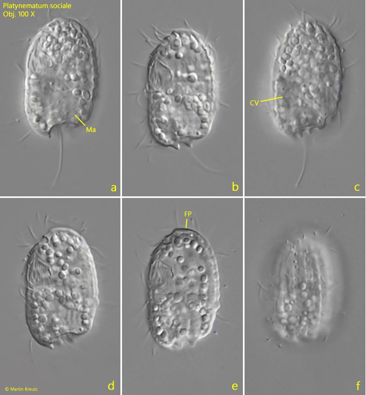
The specimens of my population were always very small, sometimes even less than 30 µm long. However, the species is easy to recognize by the ribbed pellicle and the typical notch at the posterior end, from which the caudal cilium emerges. In some specimens, the longitudinal ribs were distinctly knobby (s. fig. 1 f). The mouth opening is located almost on the narrow side of the laterally flattened body in the anterior third (s. fig. 1 a). When viewed from the left, it can be seen; from the right, it cannot. My specimens contained many highly refractive granules, which is why the position of the contractile vacuole and macronucleus was not always recognizable. In the somewhat more transparent specimens, I found the contractile vacuole shifted toward the ventral margin in the posterior third (s. fig. 2 c) and the macronucleus also in the posterior third, but shifted toward the dorsal margin (s. figs. 1 c and 2 a). The anterior end has a non-ciliated frontal plate (s. fig. 2 e).
Fig. 1 a-f:Platynematum sociale. L = 30 µm. A freely swimming specimen from left (a), right (b, c), ventral (d) and dorsal (e, f). CC = caudal cilium, Ma = macronucleus, OA = oral apparatus, PI = posterior indentation. Obj. 100 X.
Fig. 2 a-f:Platynematum sociale. L = 25 µm. A second specimen found in Nov. 2018 in the Bussenried from left. CV = contractile vacuole, FP = frontal plate, Ma = macronucleus. Obj. 100 X.