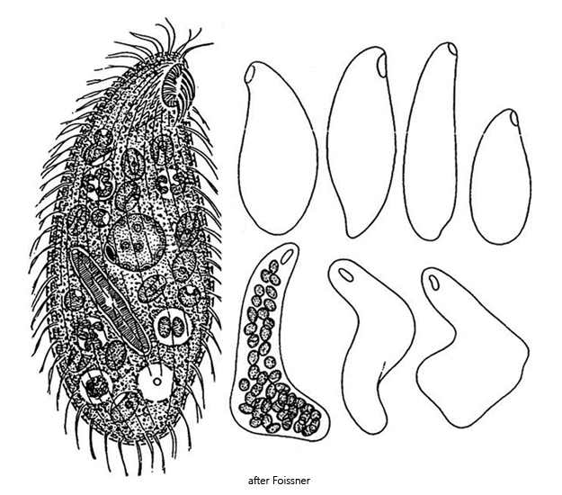
C-shaped paroral membrane right, 5–7 adoral organelles left
macronucleus spherical (9–13 µm)
micronucleus lenticular
cytoplasm green due to 30–60 symbiotic algae
symbiotic algae ellipsoid, 5–6 µm long
contractile vacuole distinctly subterminal
cortex furrowed
20–26 somatic kineties
rows of mucocysts between kineties
Platyophrya sphagni
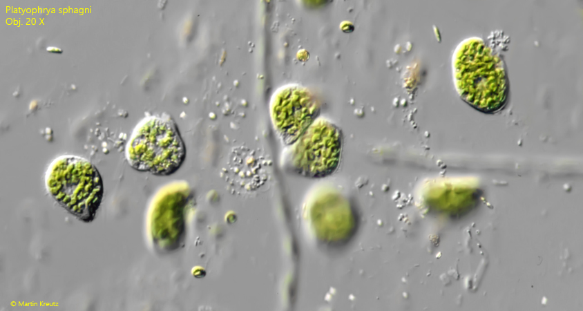
I find Platyophrya sphagni frequently and regularly in the Simmelried, but also in other bodies of water that are actually not typical because they contain no Sphagnum moss and are only slightly acidic, such as the pond at St. Ulrich. The specimens are found almost exclusively in detritus, especially when it has a gelatinous consistency and is teeming with bacteria (s. fig. 1).
Platyophrya sphagni was first described as Glaucoma sphagni by Penard in 1922. In 1993, Foissner transferred the species to the genus Platyophrya. A detailed redescription of Platyophrya sphagni was published in 1996 by Foissner & Kreutz.
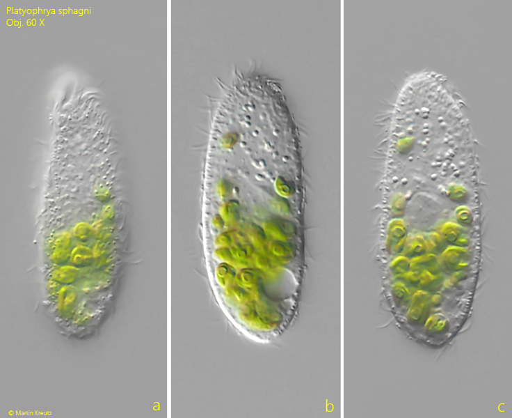
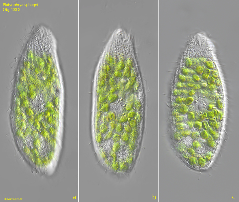
The body of Platyophrya sphagni is exceptionally capable of metaboly. When the specimens burrow through detritus flakes, they show an almost amoeboid motility. Only free-swimming specimens have an elongated, constant shape. They then usually assume a slender, pouch-like shape, with a tapered anterior end and a rounded posterior end (s. fig. 4 a-c).
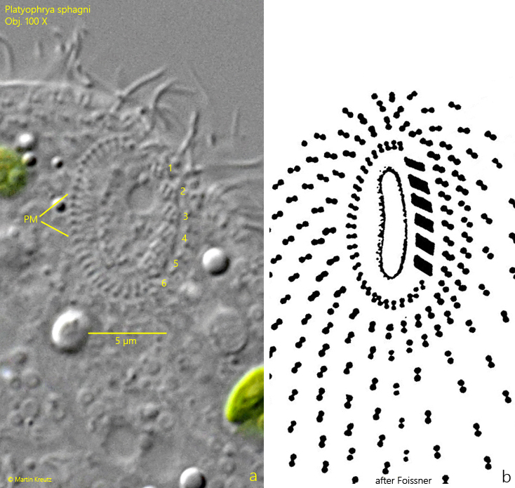
The mouth opening lies slightly subapically and is difficult to examine in motile specimens. Details can only be discerned in strongly squashed specimens that can no longer perform metaboly. A C-shaped paroral membrane is situated on the right side of the mouth opening, while an adoral zone of membranelles is situated on the left. This usually consists of 6 separate adoral organelles. This arrangement is most clearly visible when focusing on the basal bodies of the cilia (s. fig. 7 a-b).
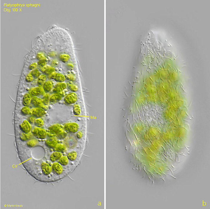
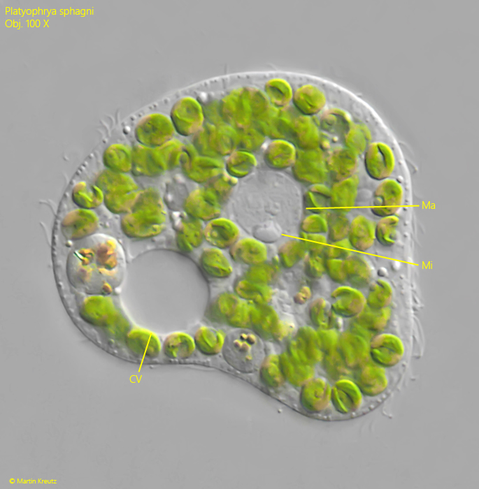
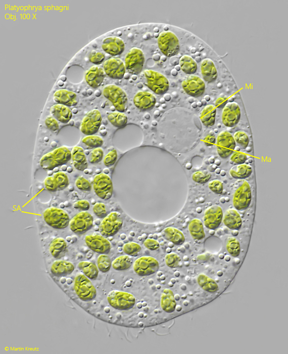
The contractile vacuole lies distinctly subterminally (s. fig. 5 a). The round macronucleus is usually found in the middle of the body. Attached to it is a lens-shaped micronucleus (s. figs. 9 and 10). There are about 20 somatic kineties, which run in a slightly spiral pattern counterclockwise (s. fig. 5 b). Between the kineties are bands of mucocysts, which are 1–2 µm long (s. fig. 8).
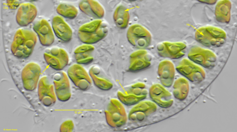
Ellipsoid-shaped zoochlorellae, which are 5–6 µm long, are always found in the cytoplasm. All cells have a pyrenoid and a nucleus (s. fig. 11). In addition, the cells usually contain an oil-like droplet. A chloroplast is present, lying against the cell wall in a plate-like shape.
Fig. 1:Platyophrya sphagni. Some specimens are rummaging through a detritus flake. Obj. 20 X.
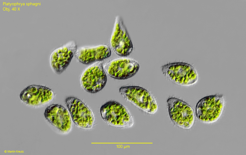
Fig. 2:Platyophrya sphagni. L = 52–71 µm. Some specimens collected from the light-exposed side of the sample vessel under the coverslip. Obj. 40 X.
Fig. 3 a-c:Platyophrya sphagni. L = 52 µm. A freely swimming specimen. Obj. 60 X.
Fig. 4 a-c:Platyophrya sphagni. L = 72 µm. A second freely swimming specimen. Obj. 100 X.
Fig. 5 a-b:Platyophrya sphagni. L = 64 µm. A slightly squashed specimen with focal plane on the spherical macronucleus (Ma) and on the somatic kineties (b). CV = subterminal contractile vacuole. Obj. 100 X.
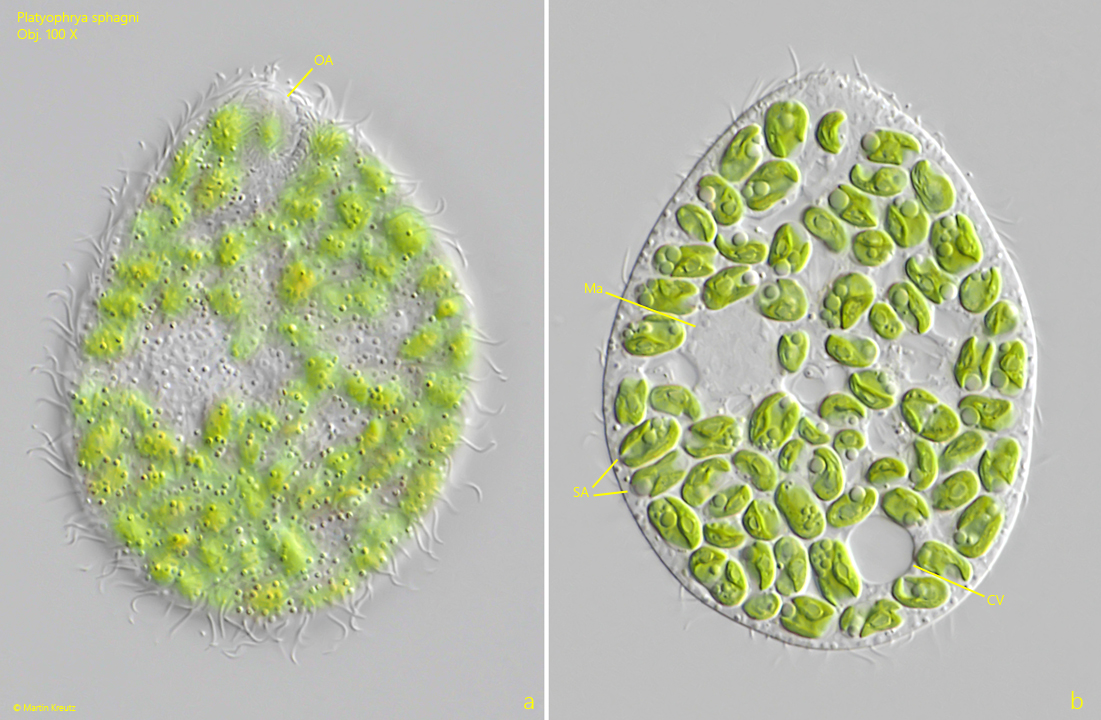
Fig. 6 a-b:Platyophrya sphagni. A squashed specimen with focal plane on the oral apparatus (OA) and the macronucleus (Ma). SA = symbiotic algae. Obj. 100 X.
Fig. 7 a-b:Platyophrya sphagni. The oral apparatus in a strongly squashed specimen (a) compared to a schematic drawing (b). The focal plane is on the basal bodies. On the right side the C-shaped paroral membrane is visible (PM) and on the left side the 6 adoral organelles. Obj. 100 X.
Fig. 8:Platyophrya sphagni. The rows of mucocysts (Mu) in a slightly squashed specimen. Obj. 100 X.
Fig. 9:Platyophrya sphagni. The macronucleus (Ma) with adjacent micronucleus (Mi) in a squashed specimen. CV = contractile vacuole. Obj. 100 X.
Fig. 10:Platyophrya sphagni. The macronucleus (Ma) and micronucleus (Mi) in a second squashed specimen. SA = symbiotic algae. Obj. 100 X.
Fig. 11:Platyophrya sphagni. The ellipsoid symbiotic algae with a length of 5–6 µm in detail. Each alga cell contain a nucleus (Nu) and a pyrenoid (PY). Obj. 100 X.