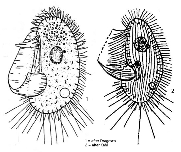
oral apparatus with large, hook-shaped unulating membrane
3 adoral membranelles (hard to see)
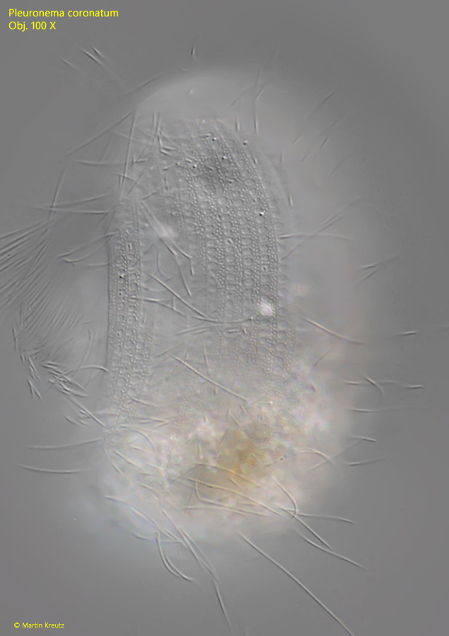
35–40 longitudinal rows of cilia
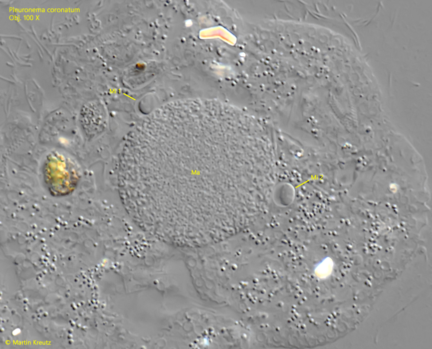
macronucleus spherical in anterior half
1–8 micronuclei adjacent to macronucleus
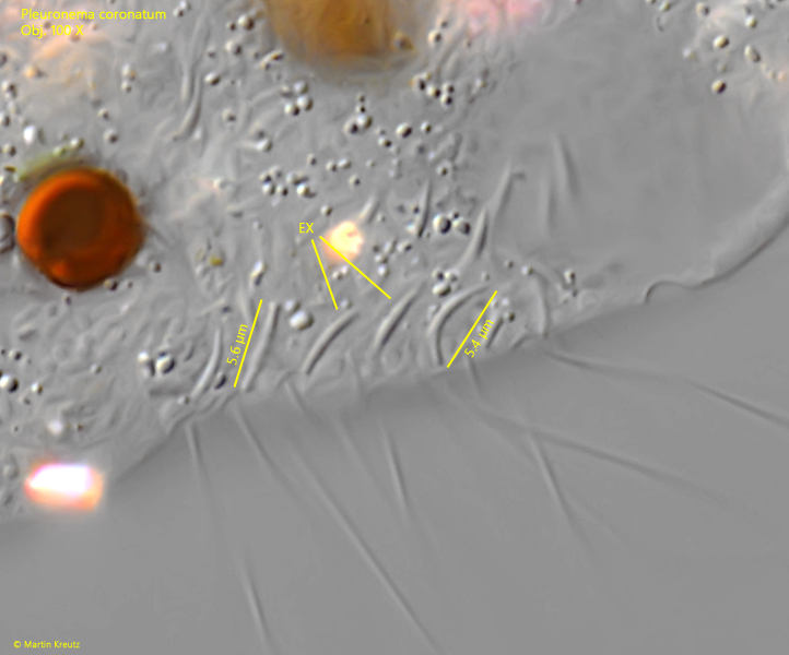
extrusomes comma-shaped, beneath pellicle
one contractile vacuole, dorsal, posterior third
posterior third with many, long caudal cilia
Pleuronema coronatum
I find Pleuronema coronatum very frequently in the top layer of mud and among decomposing plant masses in the Simmelried and Ulmisried. After placing the coverslip, the specimens initially swim very quickly but soon calm down and often remain in one spot to funnel food with the sail-shaped, large undulating membrane. Then the specimens can be well observed but react sensitively to a reduction in the layer thickness.
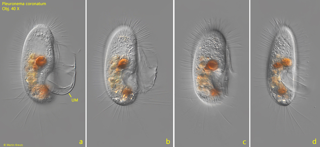
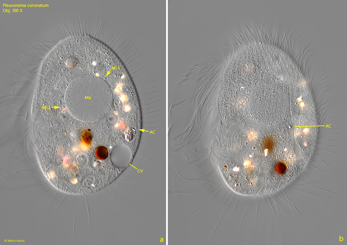
The specimens of my population were mostly between 70–90 µm long. The round, large macronucleus is clearly visible even at medium magnification (s. fig. 3). Pleuronema coronatum usually has several micronuclei. In my population, there were mostly 2–3 micronuclei present (s. figs. 3 a and 5). The most striking feature is the very large undulating membrane, which takes up about two-thirds of the body length and performs a fanning movement (s. figs. 1 a and 2 a). The exact structure of the oral apparatus is difficult to discern without silver impregnation, especially since the specimens are almost always seen only in lateral view.
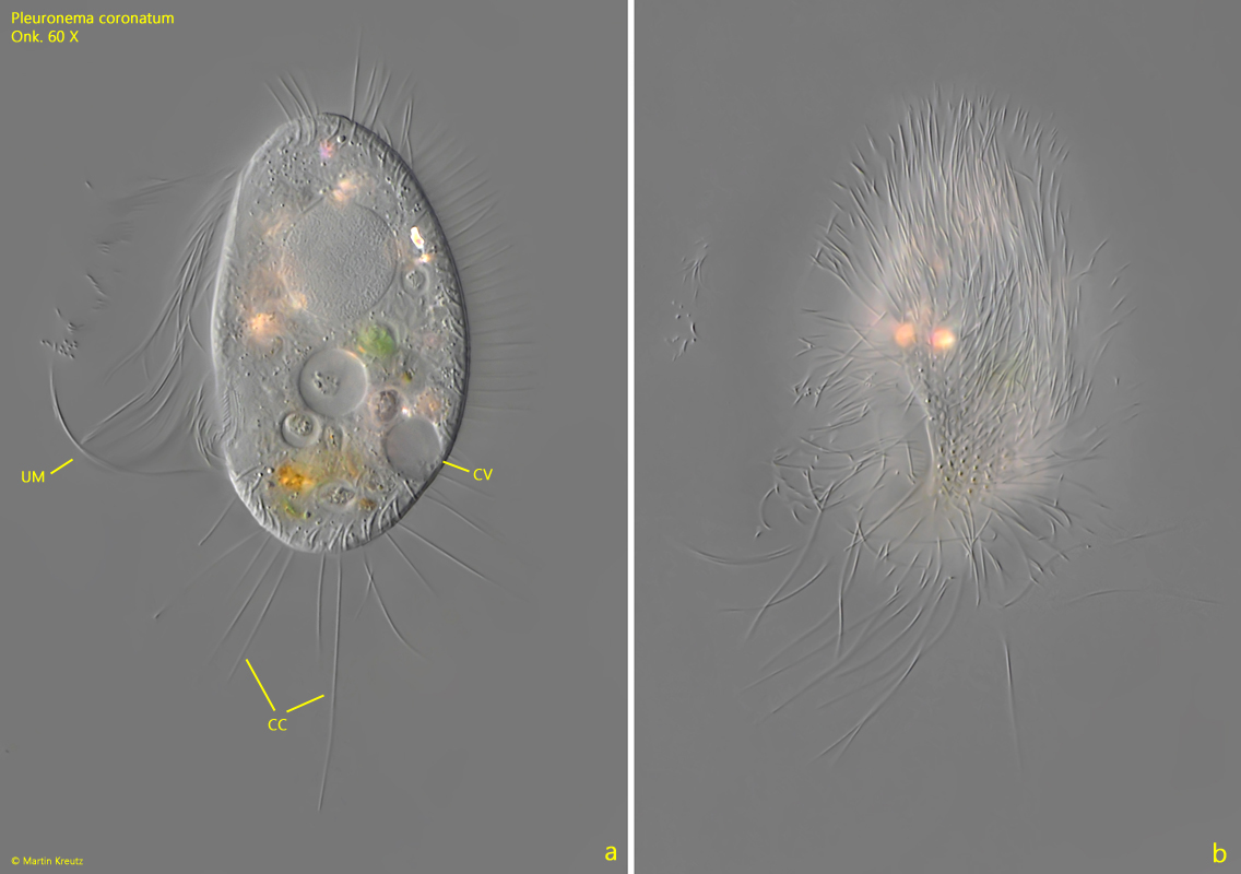
In the posterior third, there are several very long caudal cilia, which spread out fan-like in resting specimens (s. fig. 2 a). In my specimens, the middle caudal cilium was often particularly long. The contractile vacuole is located dorsally. Here, I can clearly identify an auxiliary canal that was not described by previous authors (s. fig. 3 a-b). It is consistently present, but only clearly visible in slightly squashed specimens.
When focusing on the pellicle, I was able to recognize a complicated, net-like pattern, which was also not described by previous authors (s. fig. 4). This pattern is otherwise only visible after a silver impregnation. The slightly curved, comma-shaped extrusomes of Pleuronema coronatum form a fringe under the pellicle, in which they are arranged somewhat irregularly. According to my measurements, the extrusomes are 5.4–5.6 µm long (s. fig. 6).
Fig. 1 a-d:Pleuronema coronatum. L = 95 µm. A freely swimming specimen from right (a-c) and from ventral (d). Note the large undulating membrane (UM). Obj. 40 X.
Fig. 2 a-b:Pleuronema coronatum. L = 82 µm. Two focal planes of a slightly squashed specimen from left. CC = caudal cilia, CV = contractile vacuole, UM = undulating membrane. Obj. 60 X.
Fig. 3 a-b:Pleuronema coronatum. The macronucleus (Ma) with two adjacent micronuclei (Mi 1, Mi 2). In the squashed specimen. The contractile vacuole (CV) is located dorsal in the posterior third with a single auxiliary canal (AC). Obj. 60 X.
Fig. 4:Pleuronema coronatum. L = 78 µm. Focal planes on the pellicle in a slightly squashed specimen. A complex, reticulate pattern is visible. Obj. 100 X.
Fig. 5:Pleuronema coronatum. The spherical macronucleus (Ma) with two adjacent micronuclei (Mi 1, Mi 2) in a squashed specimen. Obj. 100 X.
Fig. 6:Pleuronema coronatum. The extrusomes (EX) located beneath the pellicle are comma-shaped and have a length of 5.4–5.6 µm. Obj. 100 X.