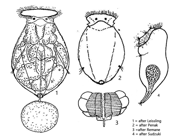
body egg-shaped, divides into four lobes in cross section
length 100–120 µm
corona oval in apical view
bordered cloacal opening at posterior end
eggs connected via thread to rotifer
two frontal eyespots
Pompholyx sulcata
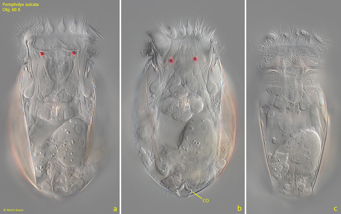
Pompholyx sulcata is one of the most common planktonic rotifers. This species often occurs in large numbers.
The characteristic features of the species is the oval body shape and two distinct eye spots located just below the corona. Pompholyx sulcata has no foot, but a clearly visible cloacal opening at the posterior end with a thickened border (s. fig. 1 b). The eggs are carried on an elastic thread and thus dragged behind while swimming. The thread, also known as “Gosse thread”, is formed by special glands located approximately in the middle of the body.
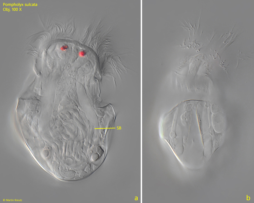
In September 2024, I observed male specimens of Pompholyx sulcata in samples from the pond of the waste disposal company Constance. Their shape did not correspond to Sudzuki’s drawing (s. drawing 4 above), which depicted the specimen naked. The male specimens in my population were 50–61 µm long and had a reduced, apparently cup-shaped lorica (s. fig. 3 a-b). The eye spots were very large and there was no digestive system. Instead, there was a large semen bladder filled with spermatozoa in the posterior half of the body (s. figs. 3 a and 4 a-b). The males are said to have an extendable, ciliated penis, but I was unable to observe this.
Fig. 1 a-c:Pompholyx sulcata. L = 107 µm. Three focal planes of a slightly squashed spcimen from ventral. Note the bordered cloacal opening (CO) at the posterior end. Obj. 60 X.
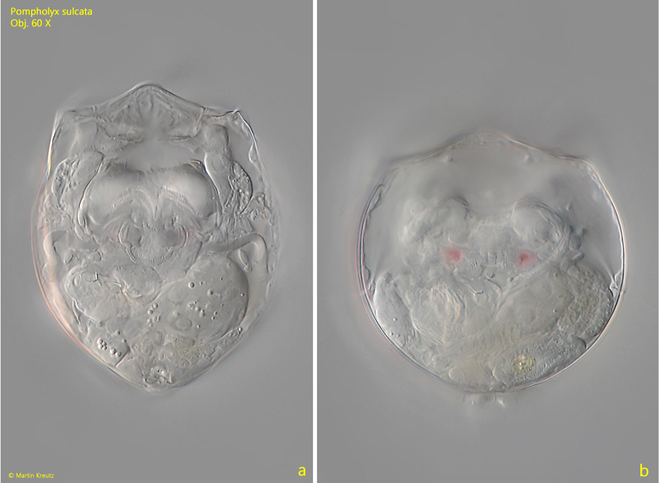
Fig. 2 a-b:Pompholyx sulcata. L = 98 µm. A partly contracted (a) and fully contracted (b) specimen from ventral. Obj. 60 X.
Fig. 3 a-b:Pompholyx sulcata. L = 57 µm. A male specimen with a reduced lorica and large eyespots. Trophi or a digestive system are not present, but a large semen bladder (SB). Obj. 100 X.
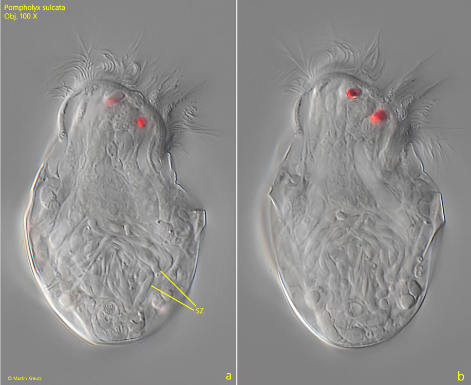
Fig. 4 a-b:Pompholyx sulcata. L = 57 µm. The slightly squashed male specimen as shown in fig. 3 a-b. In the semen bladder large spermatozoans (SZ) are visible. Obj. 100 X.