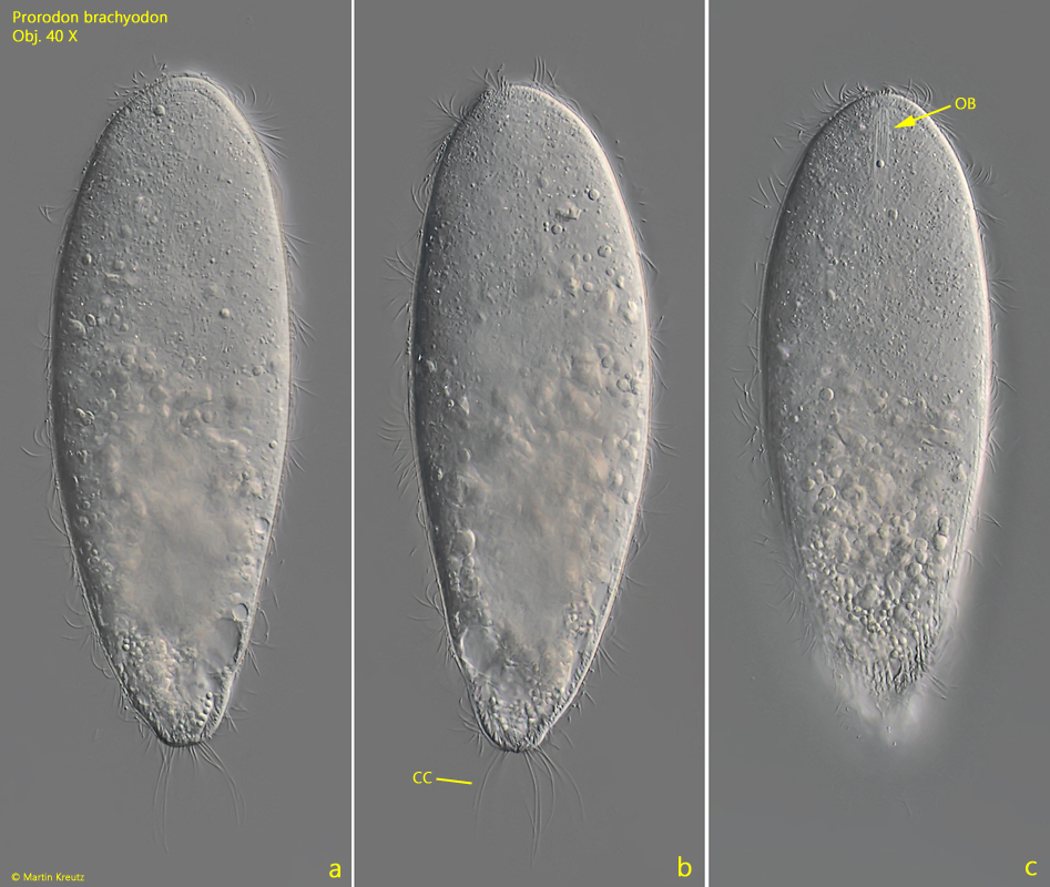
So far, I have only been able to identify a single specimen of Prorodon brachyodon, which I found in the Simmelried. The specimen was in an old sample with few plant parts.
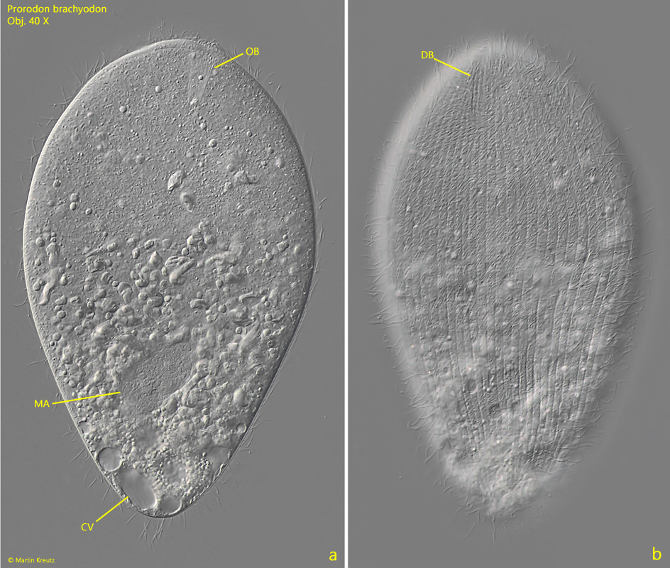
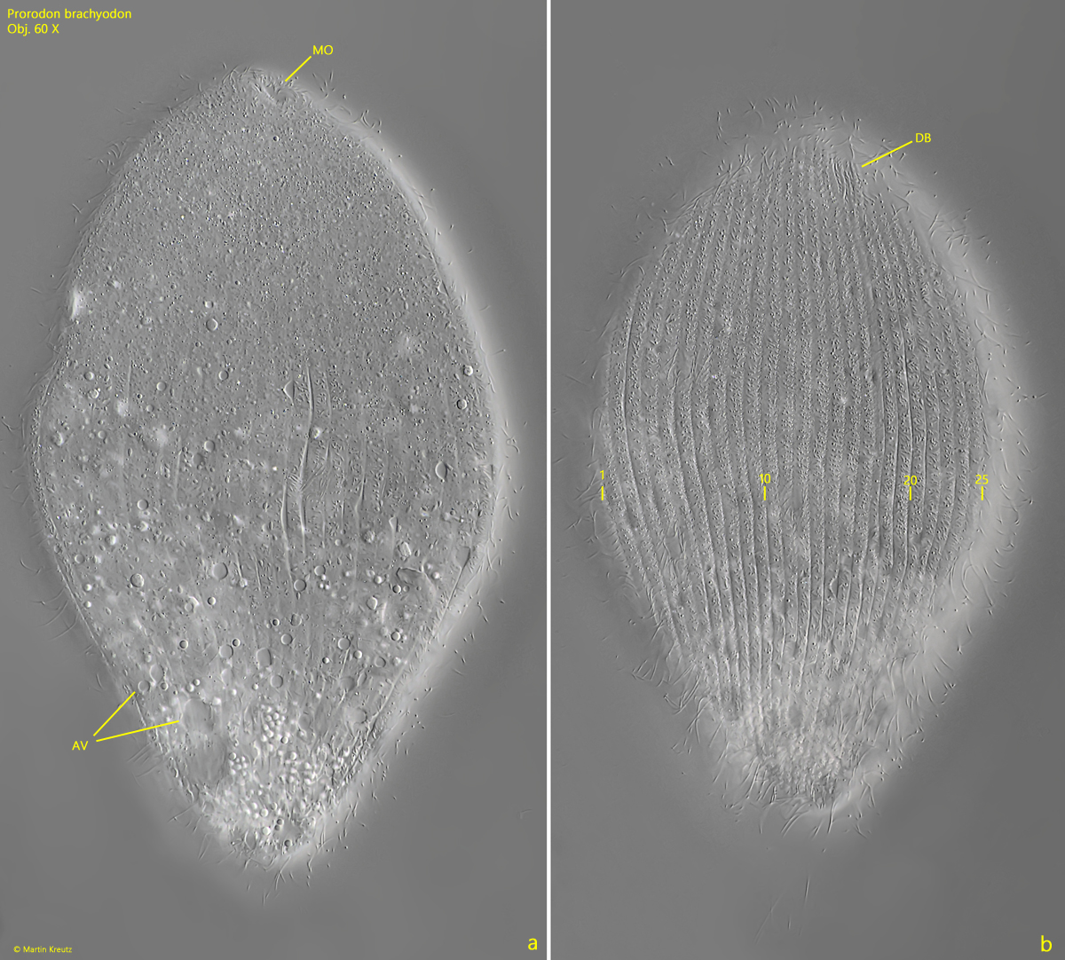
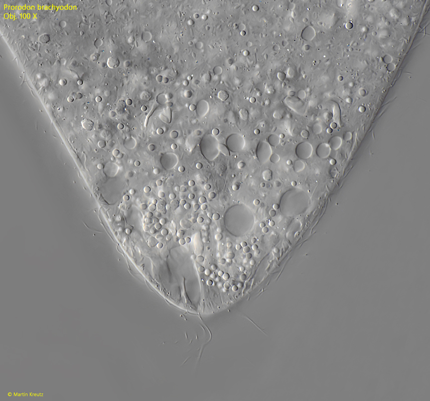
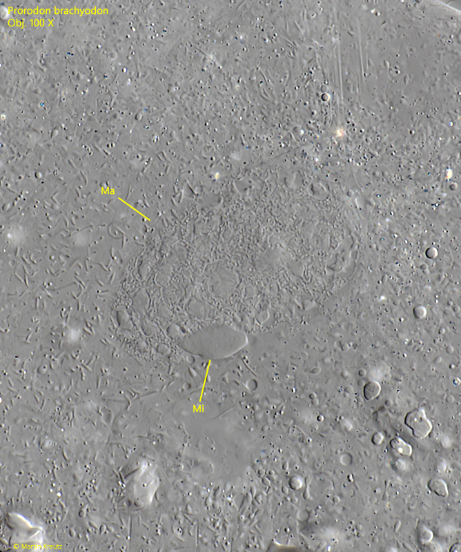
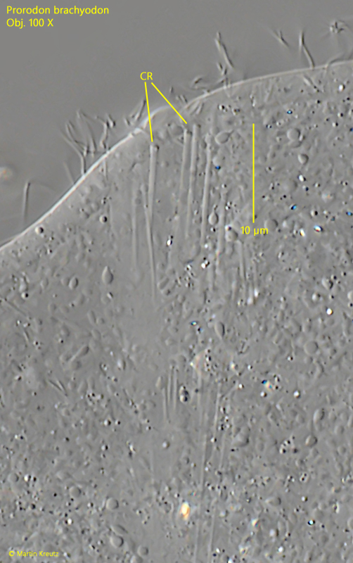
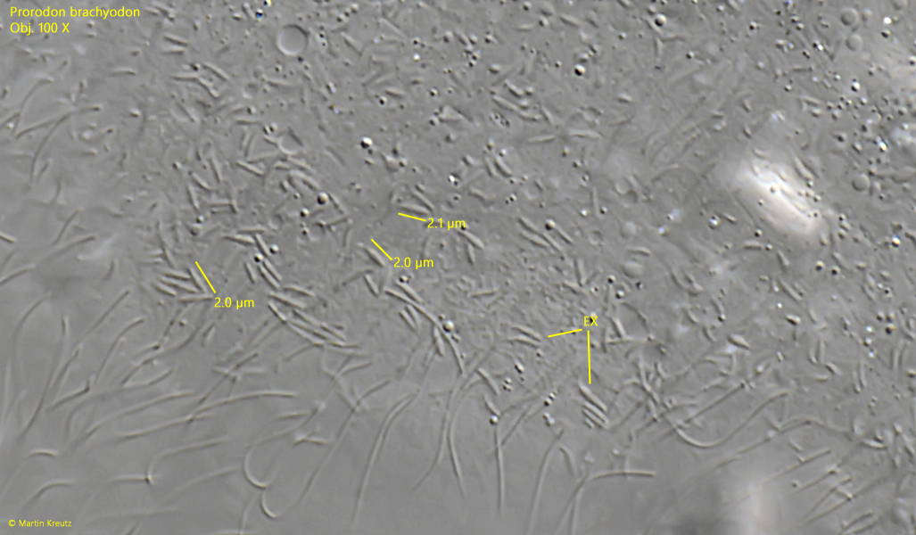
The species has so far only been briefly described by Kahl. He highlights the short oral basket as a key feature, which I was also able to confirm in my specimen (s. fig. 2 a). At the posterior end, there is a tuft of caudal cilia (s. fig. 1 b). Besides the terminal contractile vacuole, many auxiliary vacuoles can be seen just beneath the pellicle, extending about to the front third of the body. I was able to determine the number of somatic kineties to be 50–60 (s. fig. 3 b). The cytopharyngeal rods of the basket are about 10–15 µm long and nail-shaped, with a thickened head (s. fig. 6). Under the pellicle, scattered very small extrusomes can be found, which are about 2 µm long and club-shaped (s. fig. 7).
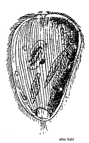
The shape of my specimen differs significantly from Kahl’s description and drawing (s. drawing above). He depicts them as very stocky. My specimen was slender and had a pointed end. Due to this deviation, I checked alternative species in the genera Holophrya and Prorodon.
Due to the distinct caudal cilia of my specimen and a length of 190 µm, many species are ruled out. The common species Holophrya teres has a similar size and shape, but a much more distinct basket and especially 80–110 somatic kineties, which is much more than my specimen.
Prorodon platyodon has a similar size to Prorodon brachyodon, up to 250 µm, but lacks auxiliary vacuoles. Additionally, Prorodon platyodon has a fringe of strong extrusomes and about 130 somatic kineties.
Other species with a length of about 200 µm and caudal cilia were not described by Kahl. I also found no further alternatives in the literature. Therefore, I believe that based on the combination of features, this is Prorodon brachyodon, even though Kahl describes and illustrates the species as stocky.