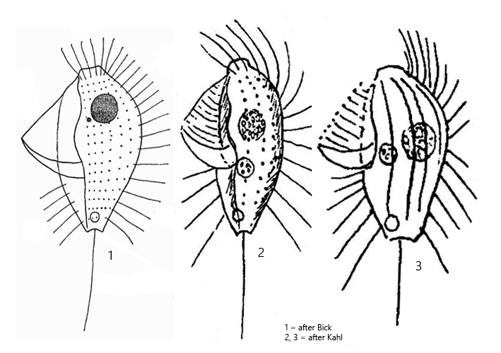
undulating membrane about two-thirds of body length
somatic cilia are paired in anterior half
posterior end truncated with a depression
one spherical macronucleus in anterior half
one spherical micronucleus in anterior end
contractile vacuole terminal
one caudal cilium
Protocyclidium citrullus
Protocyclidium citrullus was originally described by Cohn (1866) as Cyclidium citrullus. In 2002, Cyclidium citrullus was redescribed by Foissner, Agatha, and Berger. Based on morphological features of the oral apparatus (silver impregnation), the species was transferred to the genus Protocyclidium. However, a later genetic study of species within the genera Cyclidium and Protocyclidium by Gao et al. in 2014 showed that the division into the two genera is not justified. It was, however, not reversed for the time being.
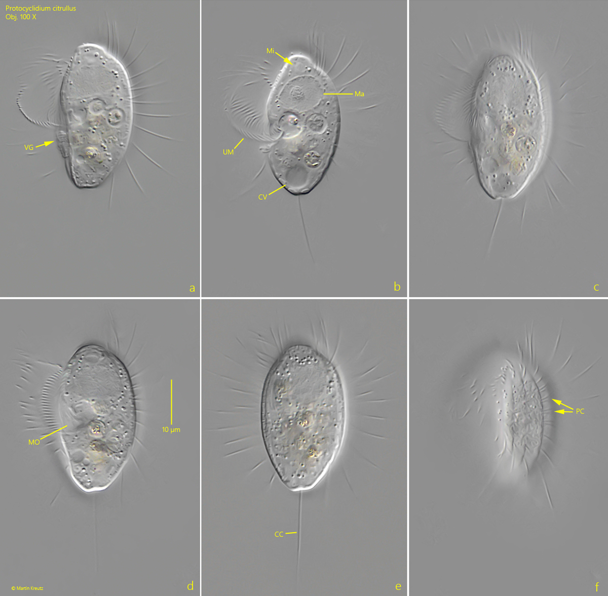
I find Protocyclidium citrullus only very rarely in my samples. Most of the time it is Cyclidium glaucoma. Protocyclidium citrullus is conspicuous because of its size (about 30% larger than Cyclidium glaucoma) and because of a ventral groove that extends to the posterior end (s. fig. 1 a). The undulating membrane is laid into it when the ciliate is at rest. This groove makes the ventral side flat, which, however, is only occasionally noticeable, depending on how the specimen is positioned in the preparation. The caudal cilium at the posterior end arises from a small depression (s. fig. 1 c). The anterior end is drawn out in a snout-like manner with a frontal plate. In my specimens the macronucleus and the micronucleus were always located in the anterior end (s, fig. 1 b) and not shifted so far towards the middle as Bick and Kahl have drawn them (see drawings above).
Fig. 1 a-f:Protocyclidium citrullus. L = 33 µm. Different focal planes of a freely swimming specimen. Note the ventral groove (VG, with adhering bacteria) into which the undulating membrane is placed when at rest. CC = caudal cilium, CV = contractile vacuole, Ma = macronucleus, Mi = micronucleus, MO = mouth opening, PC = paired cilia in anterior half, UM = undulating membrane. Obj. 100 X.