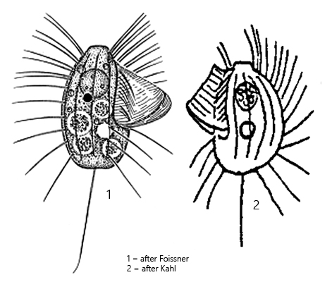
one spherical micronucleus (often near anterior end)
contractile vacuole distinctly subterminal
one caudal cilium o f body length
Protocyclidium muscicola
I found a few specimens of Protocyclidium muscicola in May 2026 in samples from the Diebelsee near Johannisthal. The Diebelsee has a bog-like character. The samples were taken from stands of Sphagnum moss and mixed with Utricularia spec.
Protocyclidium muscicola was originally described by Kahl (1931) as Cyclidium muscicola. In 1995, Cyclidium muscicola was redescribed by Foissner from soil samples taken near Salzburg and on Hawaii. Finally, based on morphological characteristics (silver impregnation), the species was transferred to the genus Protocyclidium by Foissner, Agatha and Berger in 2002.
The essential characteristics for identifying Protocyclidium muscicola are the position of the contractile vacuole just below the middle of the body and the length of the specimens of about 20 µm or less. Within the genera Cyclidium and Protocyclidium, there are only the species Cyclidium centrale and Protocyclidium terricola, which likewise possess a clearly subterminal contractile vacuole. Both species, however, are clearly larger than 30 µm.
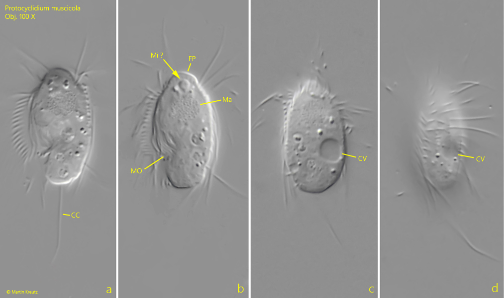
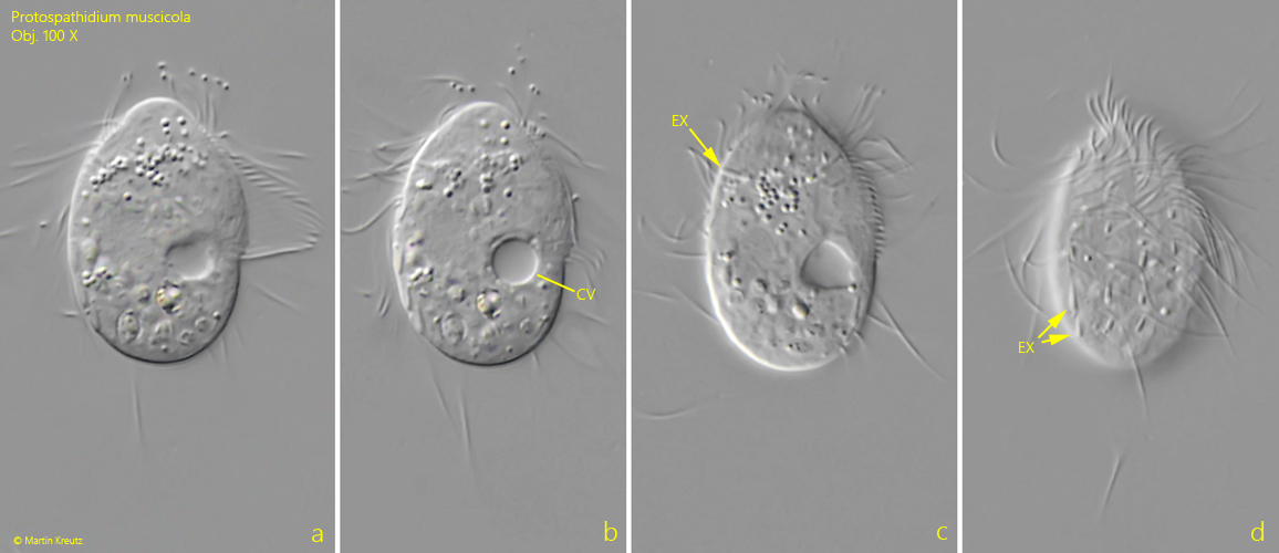
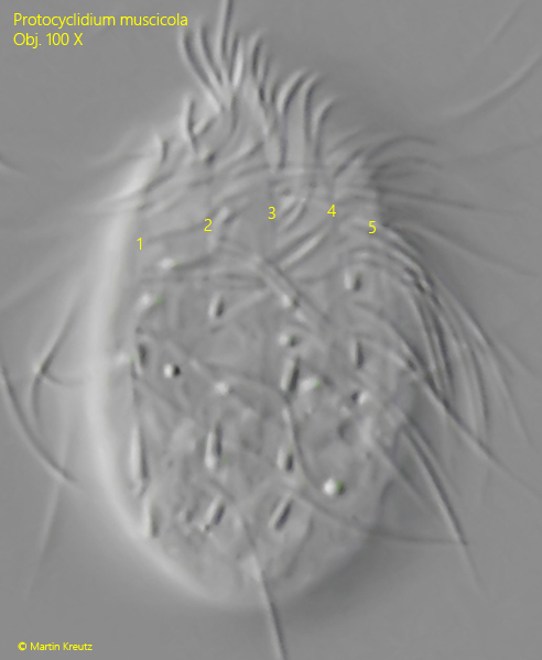
The few specimens of my population were all smaller than 20 µm. The contractile vacuole was located above the posterior third (s. figs. 1 c and 2 b). The number of kineties on the right side is 5 (s. fig. 3) what corresponds to the number of kineties found by Foissner (1995). In his redescription of Cyclidium muscicola Foissner mentions that the micronucleus is often found at the anterior end, which I was also able to recognize with high probability in one specimen (s. fig. 1b).
Deviating from the descriptions by Kahl and Foissner, I was able to observe very short, rod-shaped extrusomes in some specimens (not in all), with a length of about 2 µm (s. figs. 2 c and 2 d). The habitat also differs from the information provided by Foissner and Kahl. Foissner found populations in soil samples and Kahl in moss samples. The fact that my population was present in a moor lake may indicate a high adaptability of this species.
Fig. 1 a-d:Protocyclidium muscicola. L = 18.5 µm. Different focal planes of a freely swimming specimen from left (a, b) and from right (c, d). Note the contractile vacuole (CV) below the cell equator. CC = caudal cilium, FP = frontal plate, Ma macronucleus , Mi ? = probably the micronucleus, MO = mouth opening. Obj. 100 X.
Fig. 2 a-d:Protocyclidium muscicola. L = 23 µm. A second, slightly squashed specimen from right. In this specimen some extrusomes (EX) with a length of 2.3 µm are visible. CV = contractile vacuole. Obj. 100 X.
Fig. 3:Protocyclidium muscicola. The enlarged fig. 2 d for vizualisation of the somatic kineties. On the right side 5 kineties are visible (1-5). Obj. 100 X.