I frequently and regularly find
Pseudanabaena amphigranulata in samples from the
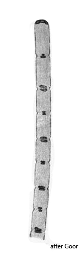
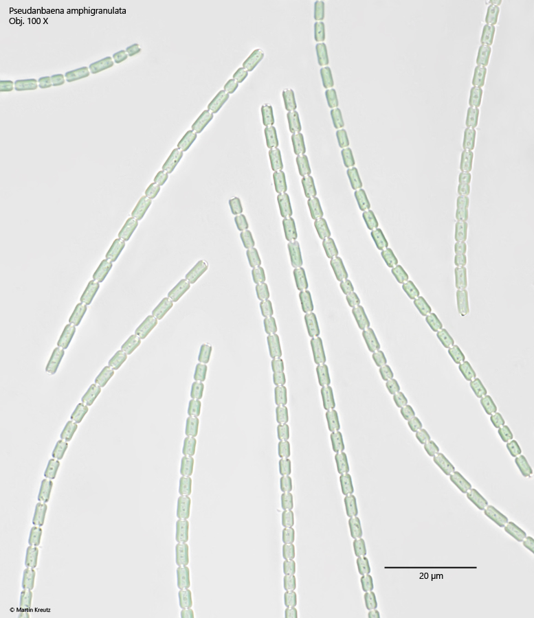
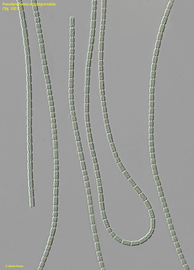
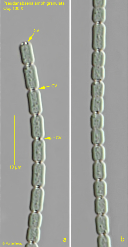
pond of the convent Hegne. The species is easy to recognize by the barrel-shaped cells with distinct constrictions at the cross walls. On both sides of the cross walls, there are small gas vacuoles (usually 2), which appear as bright spots under bright-field illumination (s. fig. 1). These gas vacuoles are also present in the apices of the terminal cells (s. fig. 3 a). The cytoplasm of the cell is divided into a lighter centroplasm and a darker stained chromatoplasm, with the chromatoplasm adjacent to the cell wall (s. figs. 1 and 3 a-b). The filaments are motile and gather after a few days on the side facing the light in the sample vessel.