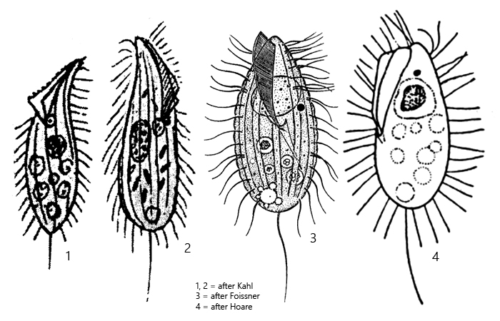
body slender ovoid, posterior rounded, anterior end narrow
length 25–50 µm, width 10–20 µm
oral apparatus with unulating membrane and adorale membrane
membranes build parallel double membrane
macronucleus spherical, in anterior third or center
one spherical micronucleus
contractile vacuole almost terminal
extrusomes absent
8–11 longitudinal ciliary rows
one caudal cilium
rapid movement
Pseudocohnilembus pusillus
So far I have only found Pseudocohnilembus pusillus in the pond at the waste disposal company Constance. However, only a few specimens. In very strongly eutrophic habitats, such as slurry puddles, this ciliate can occur in large numbers, as it is adapted to such environments.
This ciliate can be recognized at lower magnification by the rather pointed anterior end and its very rapid movement. Therefore, free-swimming specimens of Pseudocohnilembus pusillus are difficult subjects to photograph. Only in free-swimming specimens, however, does the natural, elongated shape with the narrow, drawn-out anterior end appear. When the layer thickness under the coverslip is reduced, Pseudocohnilembus pusillus quickly becomes rounded and eventually disintegrates.
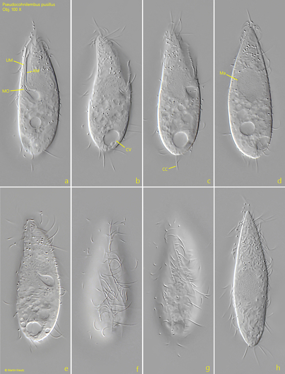
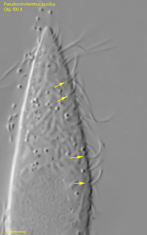
A key feature of Pseudocohnilembus pusillus is the structure of the oral apparatus. At the right margin of a groove that leads to the mouth opening in the middle of the body there is an undulating membrane. Parallel to it, to the left, there is an adoral membrane. This consists of two sections, which however cannot be discerned under a light microscope. Both membranes run parallel up to the mouth opening (s. figs. 1 a and 2 a-b). The contractile vacuole is somewhat asymmetrical and almost at the posterior end (s. fig. 1 b). At the posterior end there is a caudal cilium, which is not very long, however (s. fig. 1 c). In my specimens the macronucleus was elliptical and showed low contrast. It was located just above the middle of the body (s. fig. 1 d). I was unable to identify the micronucleus. The somatic cilia arise from small depressions (s. fig. 3), as described by Kahl (as Lembus pusillus).
Fig. 1 a-h:Pseudocohnilembus pusillus. L = 44 µm (elongated). A freely swimming specimen. Note the parallel running undulation membrane (UM) and adorale membrane (AM) of the oral apparatus and the caudal cilium (CC). CV = contractile vacuole, Ma = macronucleus, MO = mouth opening. Obj. 100 X.
Fig. 2:Pseudocohnilembus pusillus. The oral apparatus possesses a right-hand undulating membrane (UM) and an adoral membrane (AM), which consists of two parts. Both membranes lie close together and run parallel as far as the mouth opening in the center of the body. Obj. 100 X.
Fig. 3:Pseudocohnilembus pusillus. The cilia arise from small depressions (arrows). Obj. 100 X.