on left side of mouth opening 3 adoral membranelles
cyrthopharyngeal basket of 20 rods
macronucleus ellipsoidal in mid-body
contractile vacuole in mid-body
extrusomes spindle shaped
12 longitudinal rows of cilia in deep furrows
between cilia transversely striated ribs
Pseudomicrothorax agilis
So far I have only found Pseudomicrothorax agilis twice. The first time in Spechtensee in Austria in May 1999 and the second time 24 years later in July 2023 in Ulmisried. In both cases I only found a few specimens.
The specimens of my population were quite small with a length of maximum 38 µm what is at the lower limit of the range of 30–70 µm given by Foissner et al. (1994). The species is said to be widespread, but always sparse.
The food consists mainly of filamentous cyanobacteria, which are phagocytized with the cyrthopharyngeal basket (s. fig. 1 b). Unfortunately, I was unable to observe this process. The macronucleus of my specimens was spherical and located approximately in the middle of the body (s. figs. 2 a and 3). The micronucleus only becomes visible in strongly squashed specimens (s. fig. 3). The contractile vacuole is located slightly above the center of the body (s. fig. 1 c). The spindle-shaped extrusomes are about 4 µm long and arranged along the longitudinal ribs (s. fig. 2 b).
Fig. 1 a-c:Pseudomicrothorax agilis. L = 38 µm. Different focal planes of a freely swimming specimen from right. CB = cyrthopharyngeal basket, CV = contractile vacuole, Ma = macronucleus. Obj. 100 X.
Fig. 2 a-b:Pseudomicrothorax agilis. L = 32 µm. Two focal planes of a squashed specimen from right. Note the spindle-shaped extrusomes (EX) with a length of about 4 µm. CV = contractile vacuole, Ma = macronucleus. Obj. 100 X.
Fig. 3:Pseudomicrothorax agilis. The macronucleus (Ma) and micronucleus (Mi) in a strongly squashed specimen. CV = contractile vacuole. Obj. 100 X.
Fig. 4:Pseudomicrothorax agilis. L = 38 µm. Focal plane on the longitudinal ribs with a transverse striation. Obj. 100 X.
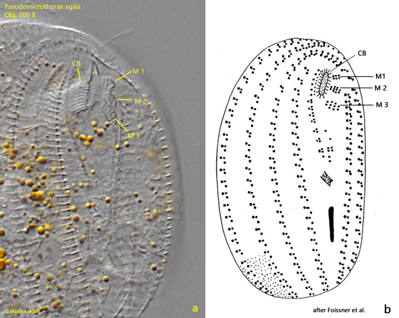
Fig. 5 a-b:Pseudomicrothorax agilis. L = 30 µm. Details of the oral apparatus in comparison with a drawing from Foissner (b). On the left side of the cyrthopharyngeal basket (CB) the three adoral membranelles (M 1, M 2 and M 3) are visible. Obj. 100 X.