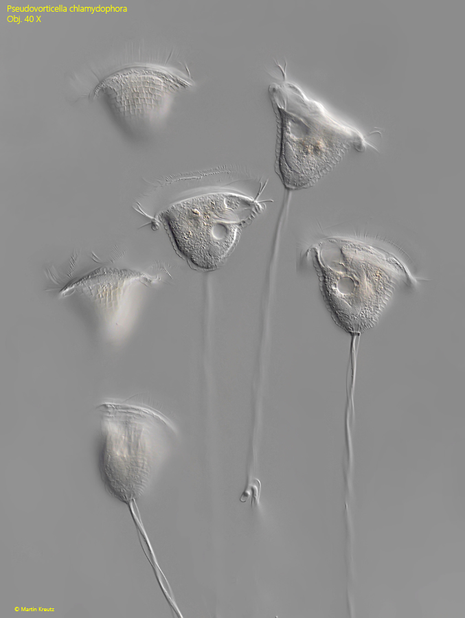
In October 2026 I found Pseudovorticella chlamydophora in the pond of the waste disposal company Constance. The pond is strongly eutrophic. A stand of Yellow Water-lily (Nuphar lutea) also grows there. I placed the leaves and stems of the Water-Lily in Petri dishes and laid floating coverslips on them. On some of these coverslips I was able to find pseudocolonies of Pseudovorticella chlamydophora after 2 days.
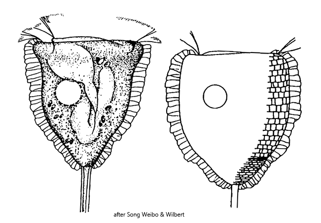
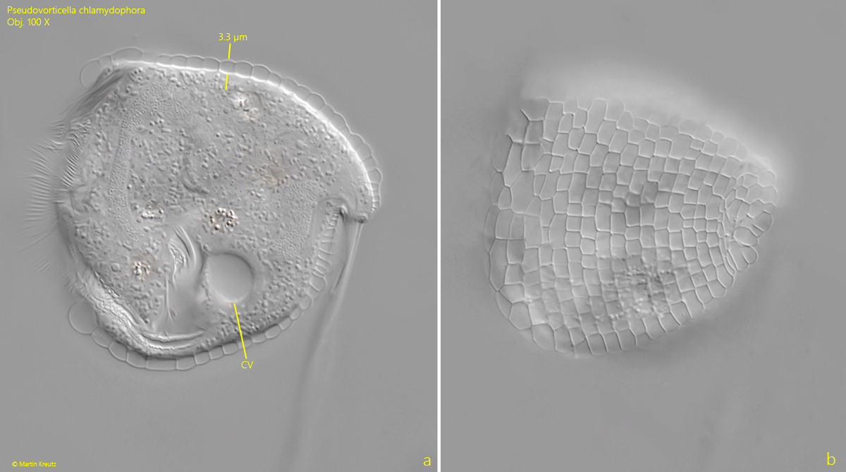
Pseudovorticella chlamydophora is a rather rare species and there are only a few records of findings. The species is very easy to recognize by the thick layer of blisters, which completely and seamlessly covers the entire surface of the zooids. These form a honeycomb-like pattern (s. figs. 3 a-b and 4). In my population the layer of blisters was 3–4 µm thick (s. fig. 3 a). Another important characteristic is the position of the contractile vacuole. Pseudovorticella chlamydophora has only one contractile vacuole, and it lies approximately at the level of the cell equator (s. fig. 3 a). The macronucleus is J-shaped. I was able to detect a single, round micronucleus (s. fig. 5), which has not yet been described in the literature.
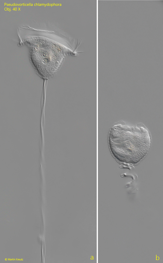
In my population the zooids had a length of 58–65 µm. The food vacuoles were mainly filled with bacteria. The contracted specimens were, in my observation, not egg-shaped, as described by Foissner, Berger & Kohmann in 1992, but rather spherical, with the layer of blisters almost closing apically (s. fig. 2 b).
Fig. 1:Pseudovorticella chlamydophora. L = 59–63 µm (of zooids). Some zooids in a pseudocolony. Obj. 40 X.
Fig. 2 a-b:Pseudovorticella chlamydophora. L = 60 µm (of extended zooid). An extended (a) and contracted zooid (b). Obj. 40 X.
Fig. 3 a-b:Pseudovorticella chlamydophora. Two focal planes of a slightly squashed specimen. The cell is covered by a layer of blisters in a reticulated pattern. In tis specimen the layer has a thickness of 3.3 µm. The single contractile vacuole (CV) is located near the cell equator. Obj. 100 X.
Fig. 4:Pseudovorticella chlamydophora. Focal plane of the reticulate pattern of the blisters in two slightly squashed zooids. Obj. 100 X.
Fig. 5:Pseudovorticella chlamydophora. The J-shaped macronucleus and the spherical micronucleus (Mi) in a squashed specimen. CV = contractile vacuole. Obj. 100 X.