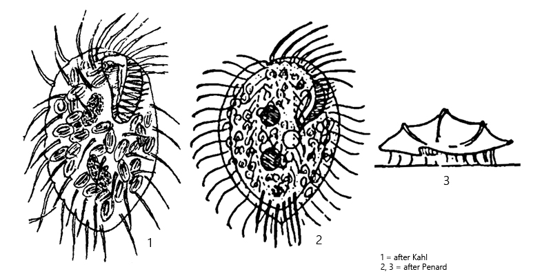
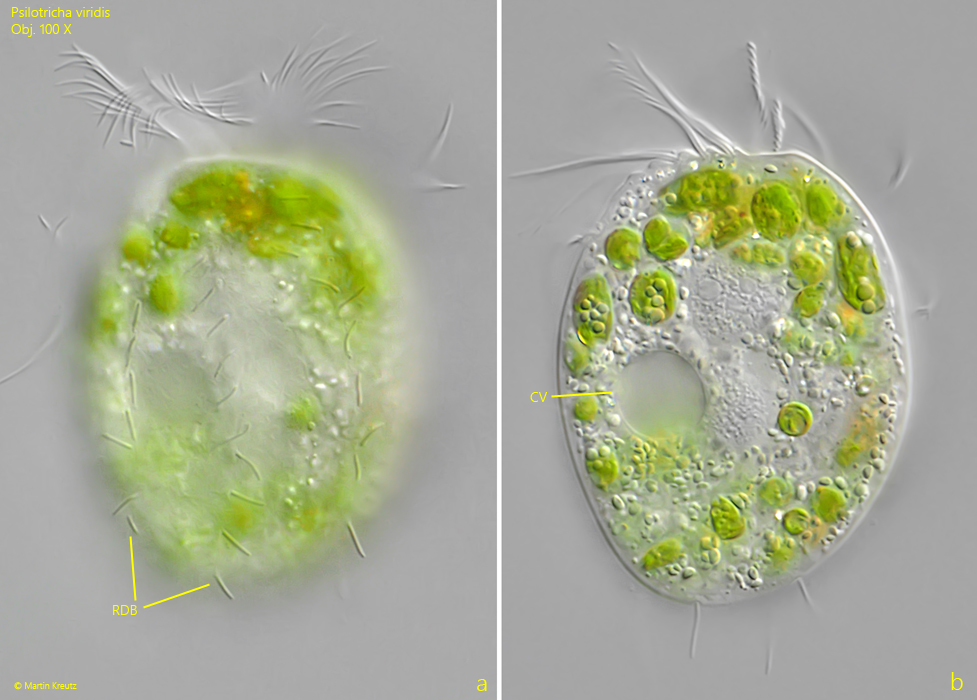
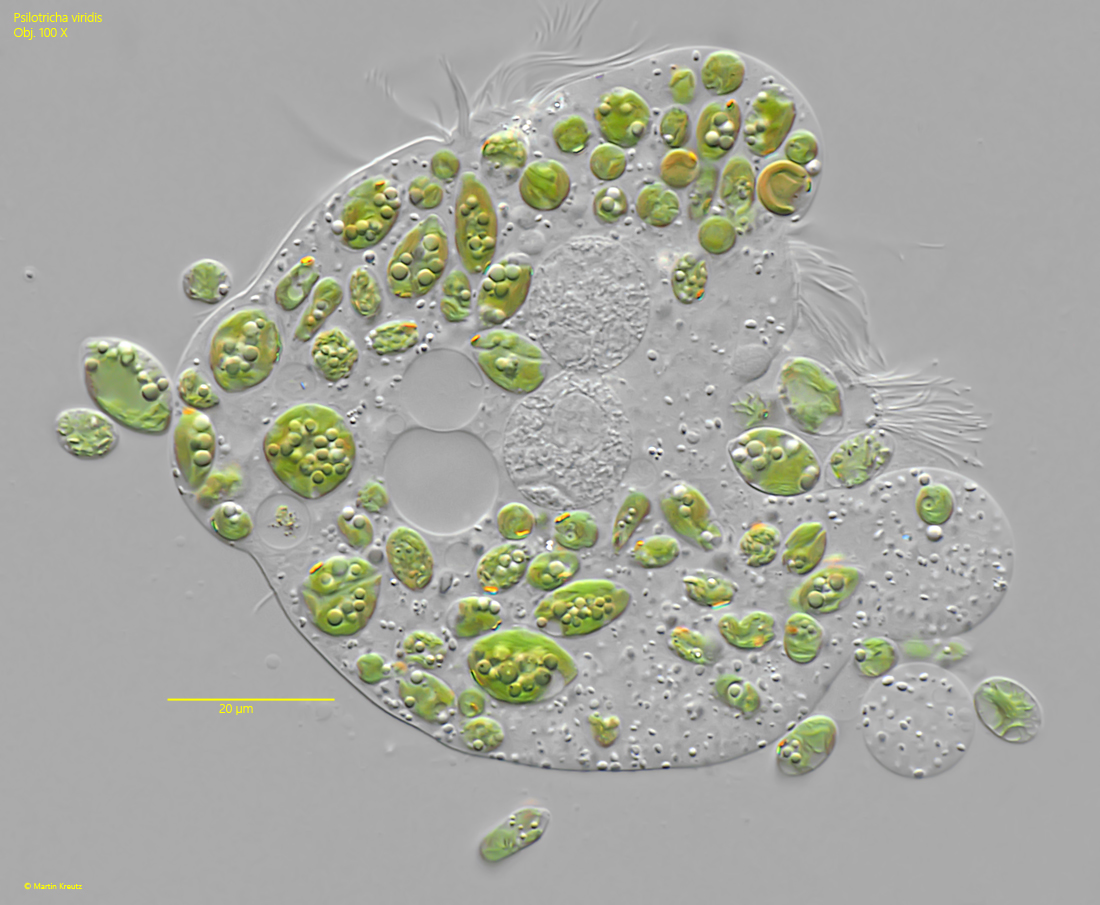
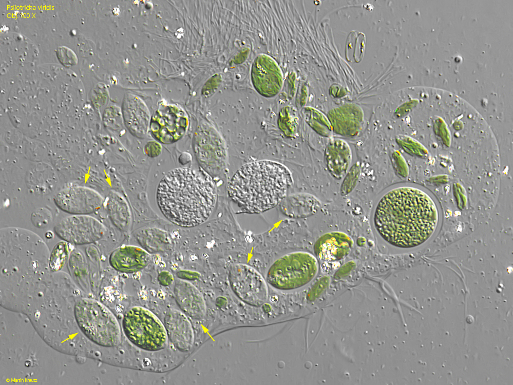
I only rarely find Psilotricha viridis and mostly in the cold season. Penard also found his specimens in January and Kahl mentions “widespread in the cold season”. The specimens shown below come from the Simmelried and from the silting-up area of the Mindelsee, where nutrient-poor puddles of meltwater form in spring. Psilotricha viridis was first assigned by Penard (1922) as Balladina viridis and transferred to the genus Psilotricha by Kahl in 1932.
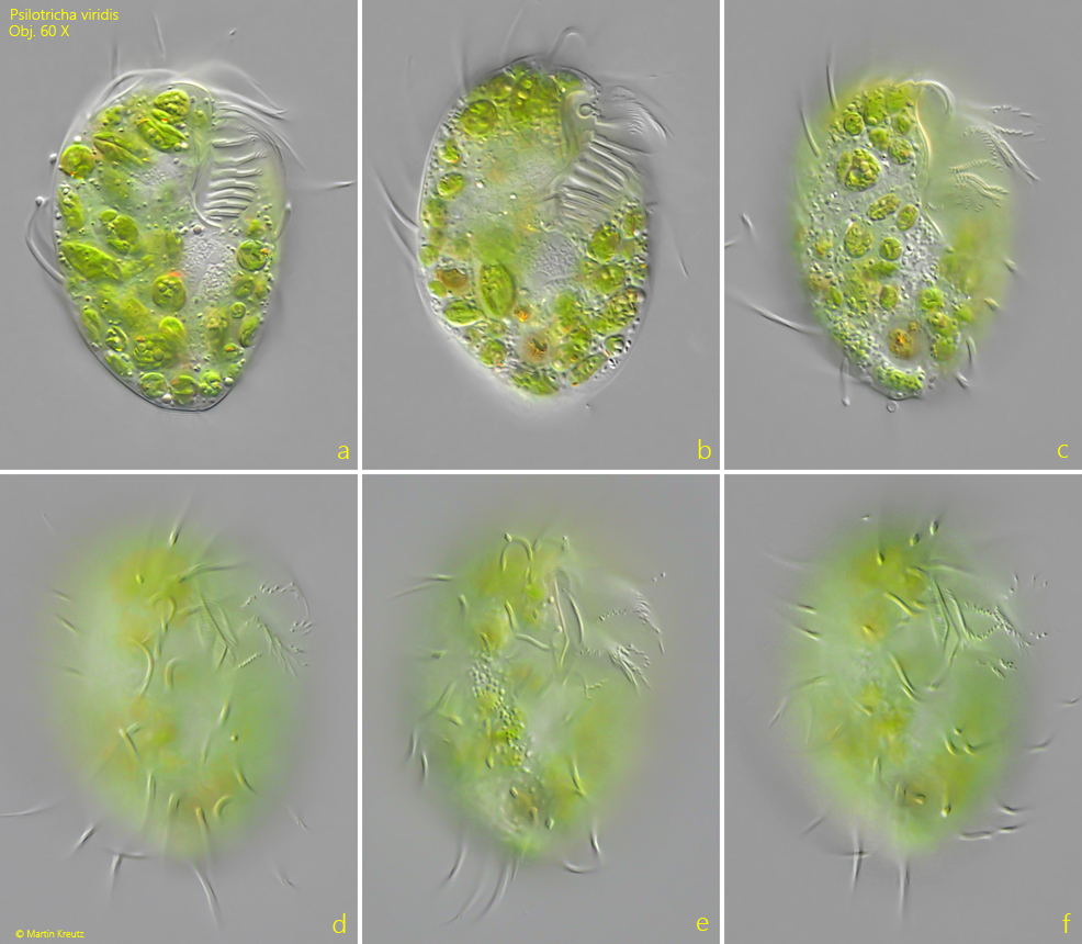
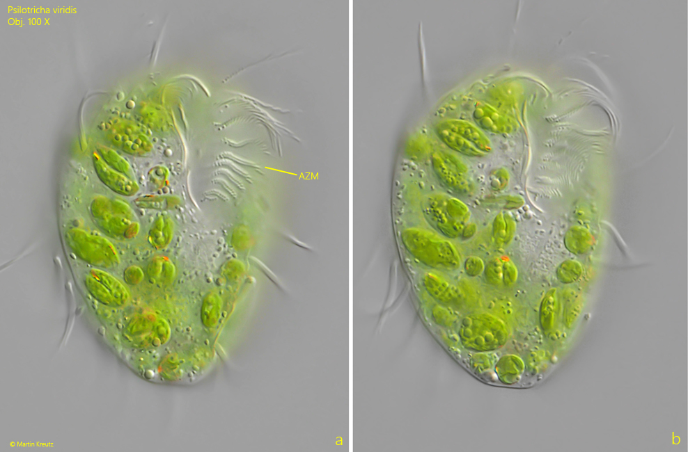
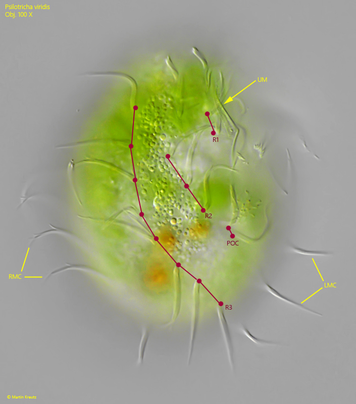
Although Psilotricha viridis is small in size, it stands out in samples due to its green coloration and turtle-like shape. On the nearly flat ventral side, the adoral zone is particularly noticeable, which extends to the first third. On the right side of the adoral zone there is an undulating membrane, which is, however, difficult to discern. The arrangement of cirri is greatly reduced in Psilotricha viridis. There are no frontal, buccal and transverse cirri. Caudal cirri are also absent. Instead, there are three rows of ventral cirri, of which the left row is the shortest and consists of only two cirri. Slightly set off below the oral apparatus are two postoral cirri (s. fig. 5). On the left and right side of the body run marginal rows of cirri.
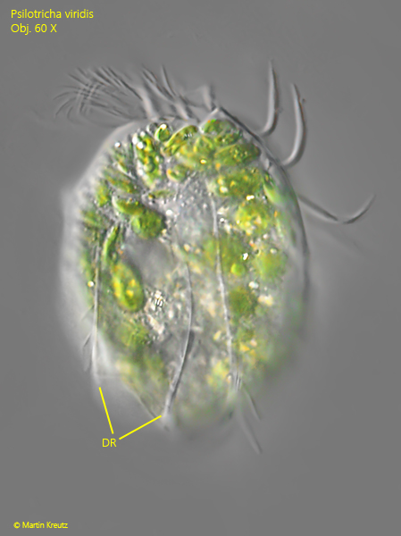
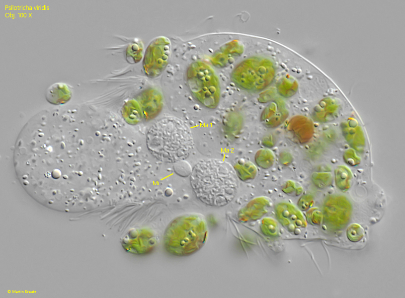
The dorsal side is slightly convex in shape and has 3 distinct ridges. Parallel to these ridges run three rows of short bristles (s. fig. 4 a-b). The contractile vacuole is also located dorsally, shifted toward the left side near the mouth opening. The nuclear apparatus consists of two spherical macronuclei, with the likewise spherical micronucleus located in their center (s. fig. 6).
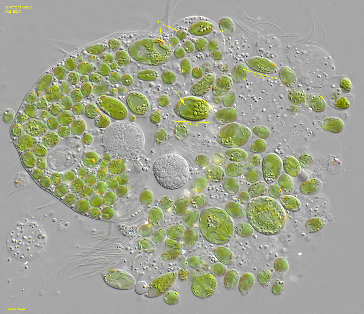
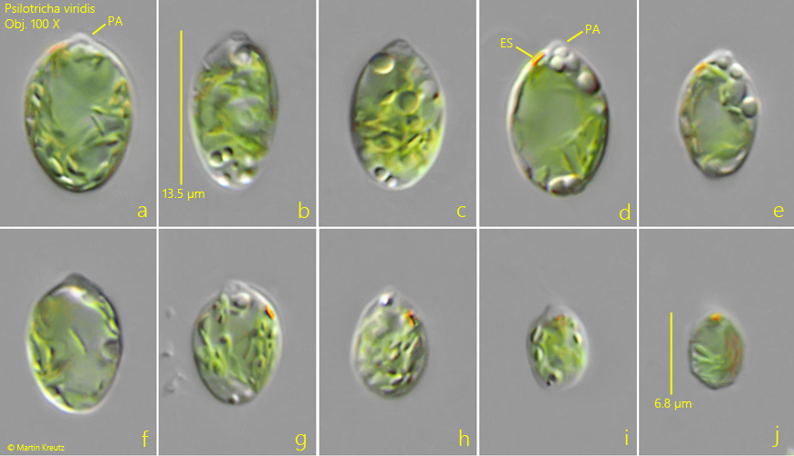
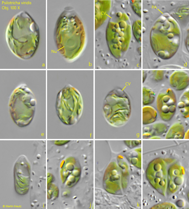
Particularly interesting are the symbiotic algae of Psilotricha viridis. They have a distinct eyespot, which is also mentioned by Kahl (1932). I was likewise able to discern this eyespot and examined the algae more closely. They are between 6–14 µm long and fully grown cells are elliptical or oval in shape. They possess their own nucleus (s. figs. 7 and 10 b), and in some cells I was also able to identify a contractile vacuole (s. figs. 10 c and 10 g). The shape of the chloroplast is hard to see. It seems to be only a single chloroplast. The algal cells often contain oily droplets. Particularly striking is a papilla at the anterior end (s. figs. 9 a, 9 d and 10 d). It is best observed when the algal cells flow out of a squashed specimen of Psilotricha viridis. This papillae and the remaining features suggest that the symbiotic algae may be members of the genus Chlamydomonas.
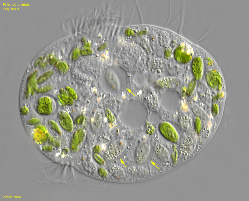
In some specimens of Psilotricha viridis I was able to find almost colorless symbiotic algae (s. figs. 11 and 12). They were obviously located in food vacuoles (s. fig. 11). This could indicate that the symbiotic algae are occasionally digested by the host cells when other food is not available or when the population of the algae becomes too large.
In 2019, Lou et al. described the similar species Hemiholosticha kahli on the island of Guam. In doing so, the authors assumed that they were examining the species Psilotricha viridis described by Kahl and, on the basis of their investigation results, concluded that this species (Psilotricha viridis sensu Kahl) must be transferred to the genus Hemiholosticha. However, the authors could only assume that it was Psilotricha viridis sensu Kahl. Proof is lacking.
Because my population matches the species Psilotricha viridis described by Kahl in all characteristics that I am able to verify, I also, with knowledge of the publication by Lou et al. (2019), continue to regard it as Psilotricha viridis.