So far, I have only found Pteromonas aequiciliata once in the plankton of the highly eutrophic pond of the waste disposal company Constance. This corresponds to the information in the literature that the species is found in nutrient-rich waters.
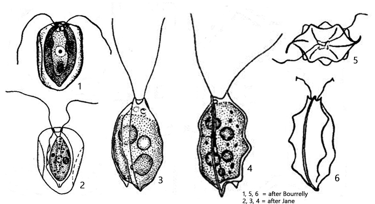
In the plankton samples, the specimens of Pteromonas aequiciliata stand out due to their abstract shape. The protoplast is surrounded by a complexly designed, transparent membrane. This membrane does not appear soft and flexible, but rather rigid and firm. The membrane has wing-like lobes and ribs, which are delicately wavy or toothed at the margins. Apically, the membrane forms two papillae through which the two equally long flagella are guided. The shape of the membranous envelope is very variable but shows recurring forms.
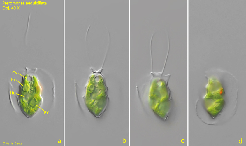
The chloroplast is said to contain 1–3 pyrenoids. I was usually able to recognize two. The protoplasts in my population were 18–29 µm long, which matches well with descriptions in the literature. The eyespot was always located laterally in the apical third. The cells swim only slowly.
Fig. 1 a-d:Pteromonas aequiciliata. L = 28 µm (of protoplast). Different focal planes of a freely swimming specimen. CV = contractile vacuole, Nu = nucleus, PY = pyrenoids. Obj. 40 X.
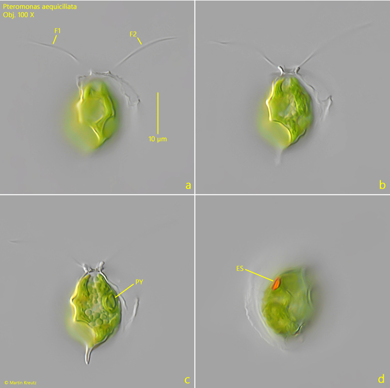
Fig. 2 a-d:Pteromonas aequiciliata. L = 19 µm (of protoplast). Different focal planes of a second freely swimming specimen. Note the two flagella (F1, F2) of equal length. ES = eyespot, PY = pyrenoid. Obj. 100 X.
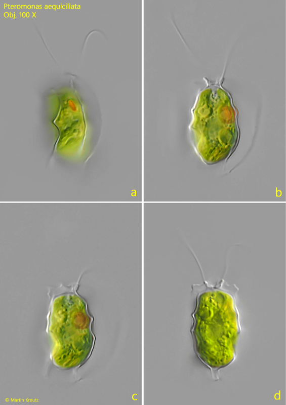
Fig. 3 a-d:Pteromonas aequiciliata. L = 21 µm (of protoplast). A third freely swimming specimen. Obj. 100 X.