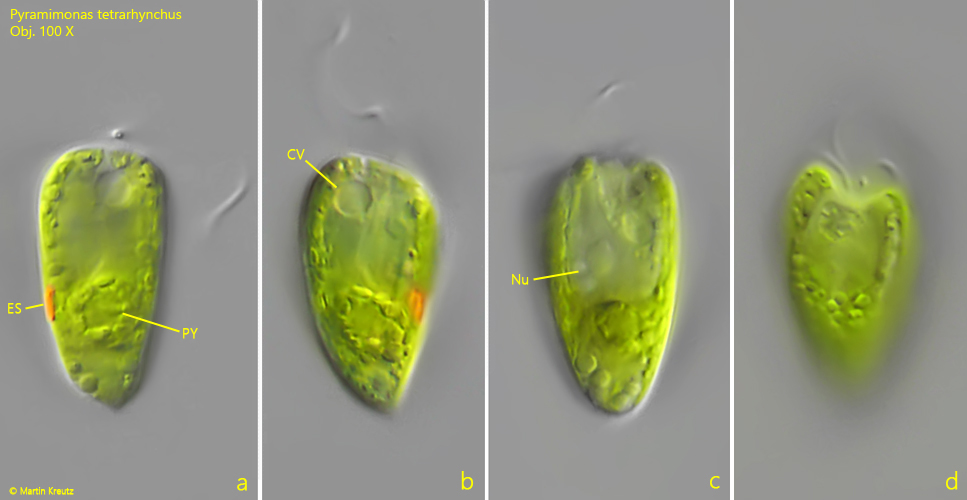
chloroplast cup-shaped, divided apically in 8 lobes
one pyrenoid, located posteriorly
one eyespot (on level of pyrenoid)
one apical contractile vacuole
Pyramimonas tetrarhynchus
So far I have only found Pyramimonas tetrarhynchus in the Simmelried, where the species rarely occurs. I usually find specimens in the spring between March and April.
The cells of Pyramimonas tetrarhynchus have 4 flagella and 4 longitudinal lobes, which are separated by furrows. This can be seen in apical view. Then the cells appear cloverleaf-shaped or square with rounded corners (s. drawing 2 above). The eyespot is clearly visible and lies in the posterior third, approximately at the same height as the pyrenoid. The cup-shaped chloroplast splits into 4 lobes, which are localized in the 4 longitudinal lobes. Apically, these 4 lobes split again to a total of 8 lobes, which is hard to recognize in living specimens.
Many authors refer to Pyramimonas rhynchomonas as Pyramidomonas rhynchomonas, for example Huber-Pestalozzi (1961).
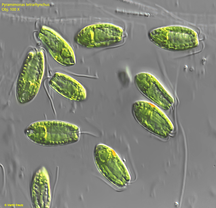
Fig. 1:Pyramimonas tetrarhynchus. L = 21–26 µm. An aggregation of several, freely swimming specimens. Obj. 100 X.
Fig. 2 a-d:Pyramimonas tetrarhynchus. L = 25 µm. Different focal planes of a freely swimming specimen. Note the 4 apical flagella and the posteriorly located eyespot (ES). Obj. 100 X.
Fig. 3 a-d:Pyramimonas tetrarhynchus. L = 26 µm. A second freely swimming specimen. Note the pyrenoid (PY) on the level of the eyespot (ES). CV = contractile vacuole, Nu = nucleus. Obj. 100 X.