I found Rhabdogloea linearis in samples from the Schwemm Moor in Austria, which were several weeks old. I found a few colonies on the walls of the sample containers.
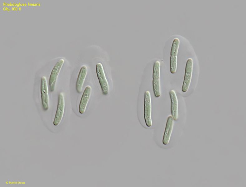
The colonies in my population consisted of 4–10 cells. The mucous sheath was clearly visible and weakly layered around the cells. The rod-shaped cells were slightly larger than those described by Komarek & Anagnostidis (1999), measuring 8–15 µm in length, but otherwise corresponded to the description. The cells were faint blue-green. I could see very small orange vesicles, especially near the cell ends (s. fig. 1).
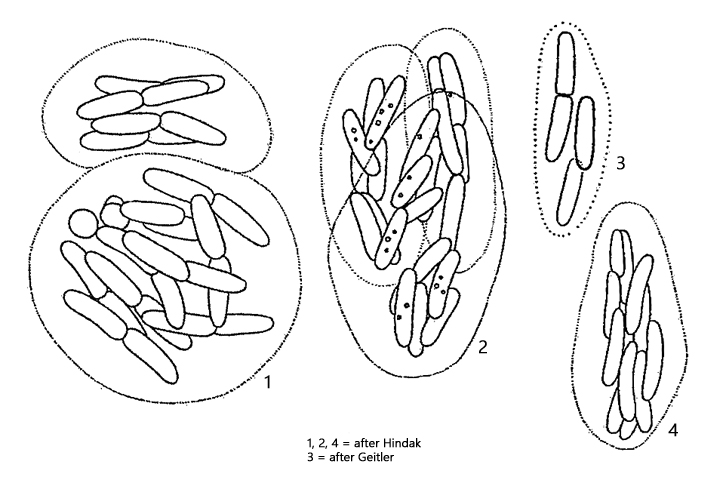
The similar species Rhabdogloea smithii has spindle-shaped cells with distinctly pointed ends.
Fig. 1:Rhabdogloea linearis. L = 11.5–14.5 µm (of cells). Two colonies of each six cells. Obj. 100 X.
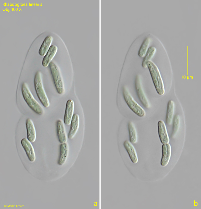
Fig. 2 a-b:Rhabdogloea linearis. L = 8.3–12.9 µm (of cells). Two focal planes of a colony with 10 cells. One cell is in the process of cell division. Obj. 100 X.