stalk not contractile, very short, sometimes absent
length 45–77 µm
peristome collar about body wide
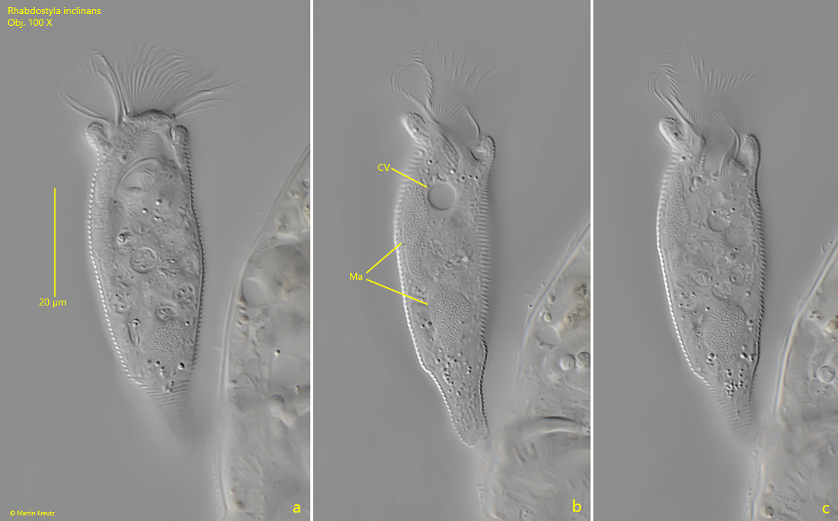
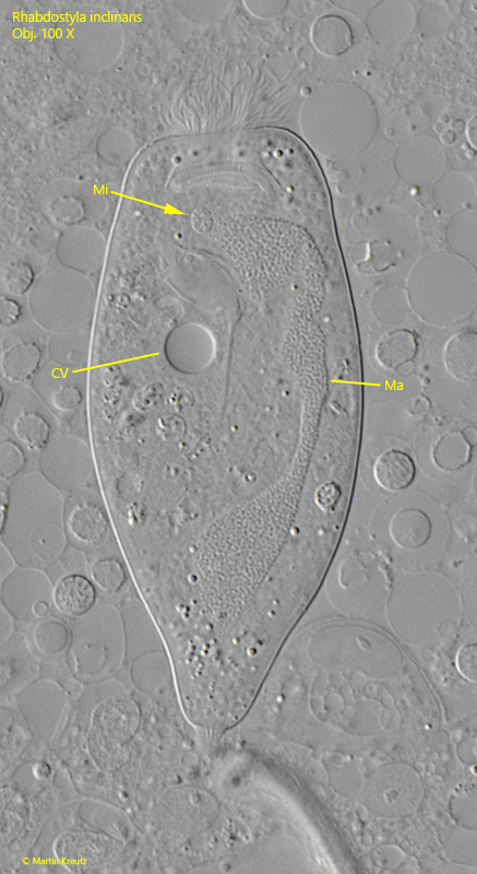
macronucleus almost body length, longitudinally arranged
one spherical micronucleus
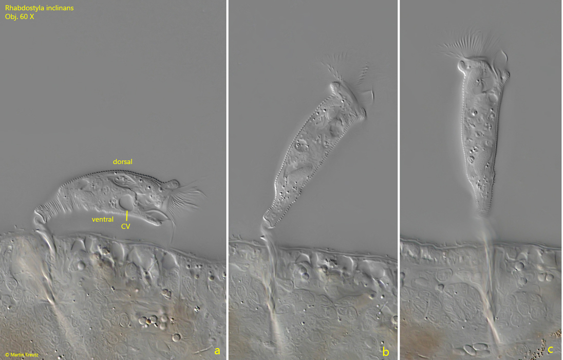
contractile vacuole in posterior third, on ventral side of oral funnel
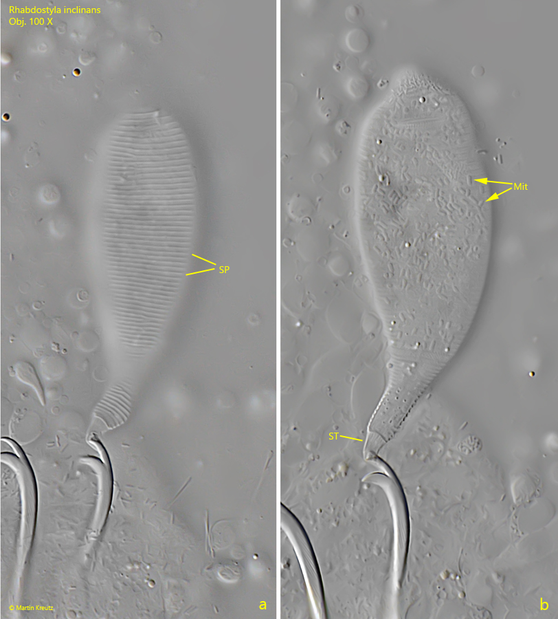
distinct striation of the pellicle
epizoic lifestyle on epidermis of oligochaetes
Rhabdostyla inclinans
I find Rhabdostyla inclinans particularly frequently in old samples where oligochaetes have multiplied and live in the sludge layer or on the walls of the sample containers.
Rhabdostyla inclinans has an epibiotic lifestyle and very often attaches itself to the bristles of oligochaetes, such as Nais species. Even at low magnification the specimens of Rhabdostyla inclinans attached to the bristles of the oligochaetes can be recognized. I have never observed that Rhabdostyla inclinans settles directly on the epidermis of the oligochaetes. Despite the movement of the oligochaetes through the sludge, the attached specimens of Rhabdostyla inclinans cannot be brushed off. Under coverslip pressure, the specimens do contract but remain attached to the bristles.
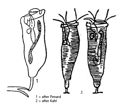
The specimens of my population were between 60–70 µm long. The body was always slender and roughly pear-shaped, with the posterior quarter of the body always distinctly tapered (s. figs. 2 b and 3 b). The stalk, with which the specimens attach themselves to the bristles, was always very short, measuring 4–6 µm in length (s. fig. 4 b). Penard (1922) reports a species Syphidia discostyla, which forms an adhesive disc without a stalk to settle directly on the epidermis of Nais spec. Kahl already suspected in 1935 that this might also be Rhabdostyla inclinans, which can also transform the stalk into an adhesive disc, and considers the two species synonymous. Foissner et al. (1992) also regard this as plausible.
The pellicle of my specimens was very distinctly transversely striated (s. fig. 4 a). The contractile vacuole is located on the ventral side adjacent to the oral funnel and is somewhat set back from the apical end (s. fig. 2 a). The extended peristome is as wide as the body or only slightly wider. The elongated macronucleus lies parallel to the longitudinal axis of the cell, and at the apical end there is a spherical macronucleus (s. fig. 5).
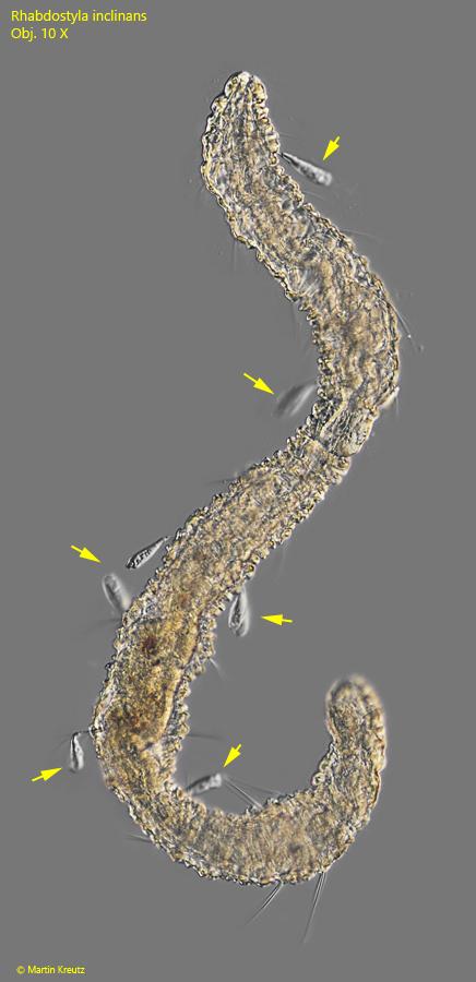
Fig. 1:Rhabdostyla inclinans. L = 180 µm. Some specimens (arrows) attached to the bristles of an oligochaete (likely Aeolosoma spec.). Obj. 10 X.
Fig. 2 a-c:Rhabdostyla inclinans. L = 62 µm. A specimen attached to the bristles of the oligochaete Nais spec. Note the contractile vacuole (CV) located on the ventral side of the oral funnel. Obj. 60 X.
Fig. 3 a-c:Rhabdostyla inclinans. L = 66 µm. A specimen attached to the bristles of the oligochaete Nais spec. in detail. The posterior part of the body is noticable tapered. The elongated macronucleus is arranged parallel to the longitudinal axis of the cell. CV = contractile vacuole. Obj. 100 X.
Fig. 4 a-b:Rhabdostyla inclinans. In a squashed specimen the distinct striation of the pellicle (SP) is visible as well as the short, inconspicuous stalk (ST). Mit = mitochondria beneath the pellicle. Obj. 100 X.
Fig. 5:Rhabdostyla inclinans. The macronucleus (Ma) and the adjacent micronucleus (Mi) in a strongly squashed specimen. CV = contractile vacuole. Obj. 100 X.