Rhizamoeba clavarioides

(Penard, 1902) Siemensma, 1980

Most likely ID: n.a.


Synonym: n.a.


Sampling location: Simmelried


Phylogenetic tree: n.a.


Diagnosis: 

  • body monopodial or polypodial
  • pseudopodia flat
  • length 70–220 µm
  • nucleus (9.6–15.5 µm) ovoid or spherical with nucleolus  
  • nucleolus irregular or clavate
  • one contractile vacuole
  • sometimes covered with debris
  • uroid bulbous with trailing uroidal filaments
Rhizamoeba-clavarioides
Rhizamoeba clavarioides
So far, I have found Rhizamoeba clavarioides exclusively in the top layer of mud in the Simmelried and only up until the year 2014. After that, I have not found any more specimens.
 
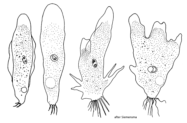
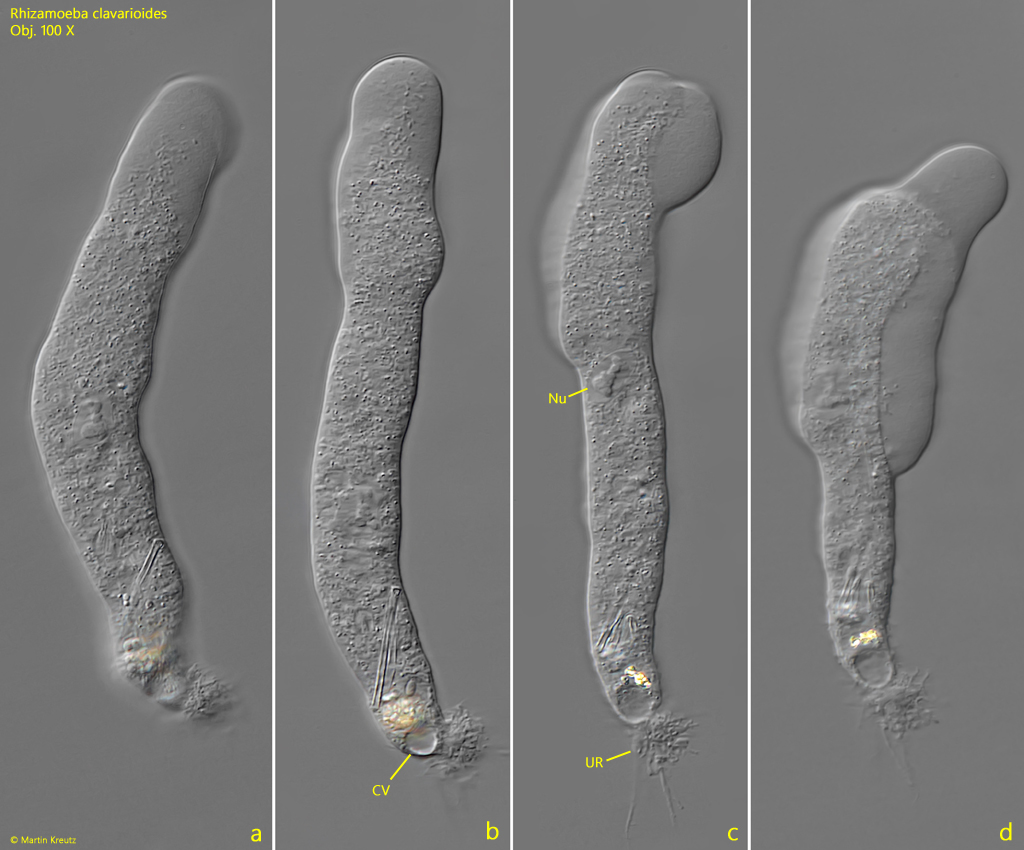
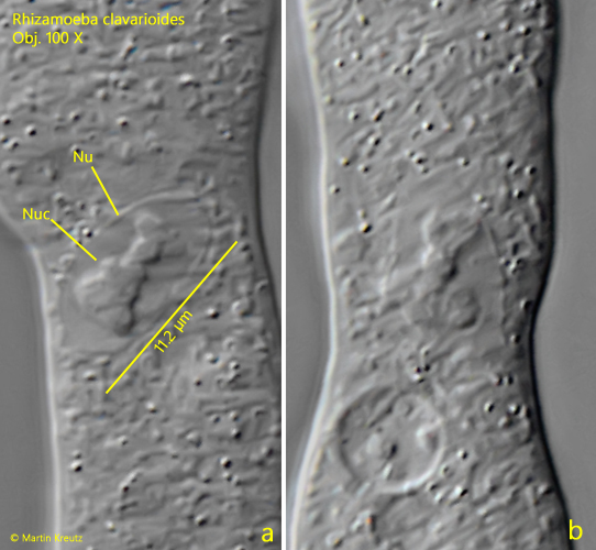
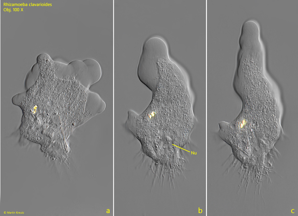
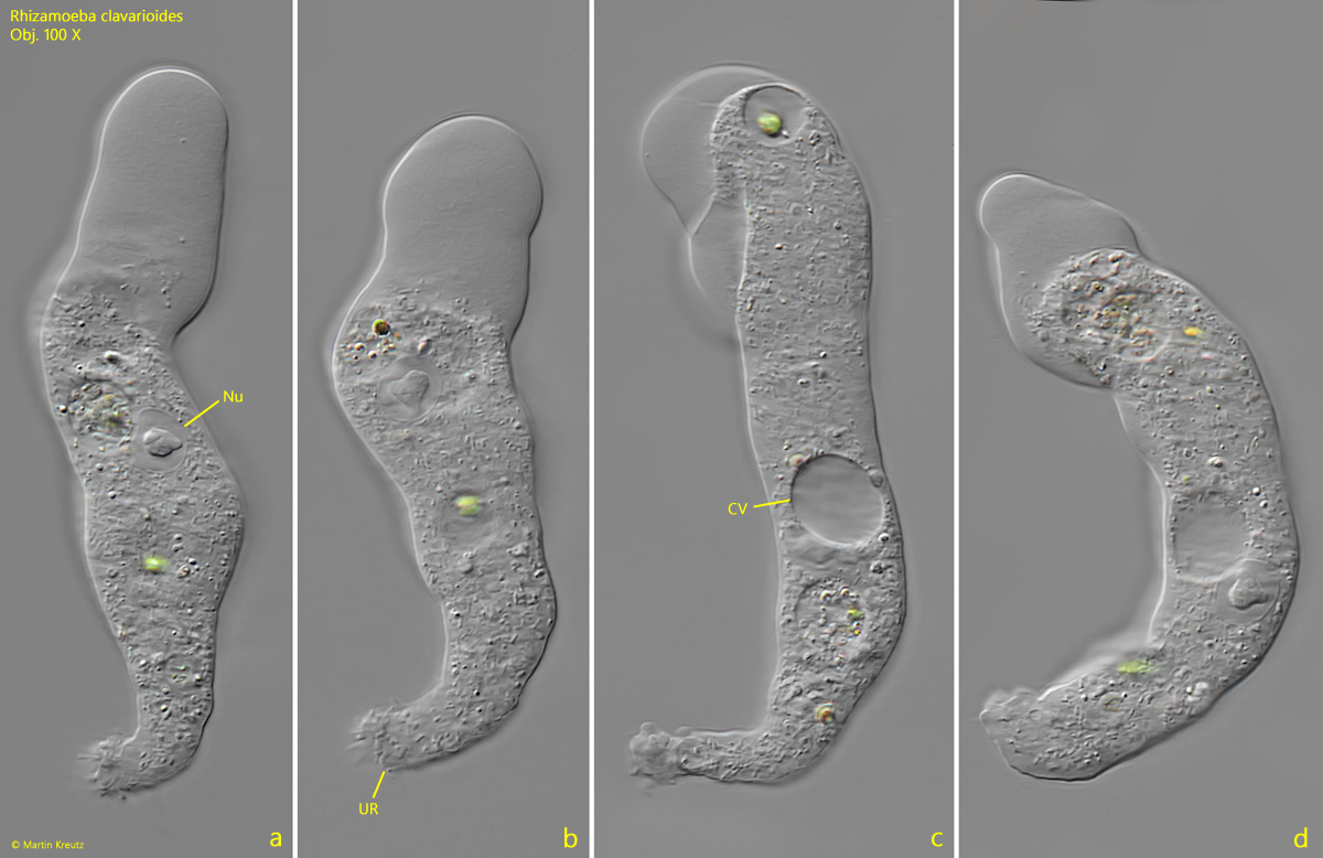
Under the coverslip, Rhizamoeba clavarioides usually takes on the monopodial limax form. In this form, movement is fast. Key features of Rhizamoeba clavarioides are the uroid, with very thin trailing filaments, and the irregularly shaped nucleolus in the oval or ellipsoid-shaped nucleus (s. figs. 1 c and 2 a-b). These features allow Rhizamoeba clavarioides to be distinguished well from other species. The similar species Rhizamoeba coerulea is larger, measuring 150–260 µm, and also has multiple nuclei with irregularly shaped nucleoli.

Sometimes specimens of Rhizamoeba covered with detritus are also found. Usually, in these specimens, the nucleus is not clearly visible and identification is difficult. However, after placing the coverslip, the specimens mostly leave this protection, allowing for closer examination.
 

More images and information on Rhizamoeba clavarioides: Ferry Siemensma-Microworld-Rhizamoeba clavarioides

Rhizamoeba-clavarioides

Fig. 1 a-d: Rhizamoeba clavarioides. L = 118 µm. Different stages of a moving specimen in the monopopdial limax form. Note the irregular shaped nucleous in the nucleus (Nu) and the uroid (UR) with thin uroidal filaments. Obj. 100 X.

Rhizamoeba-clavarioides

Fig. 2 a-b: Rhizamoeba clavarioides. The nucleus (Nu) and the irregularly shaped nucleolus (Nuc) in detail. The ellipsoid nucleus has a length of 11.2 µm. Obj. 100 X.

Rhizamoeba-clavarioides

Fig. 3 a-c: Rhizamoeba clavarioides. L = 91 µm. The same specimen as shown in fig. 1 a-d start to change from the polypodial form to the monopodial limax form. Obj. 100 X.

Rhizamoeba-clavarioides

Fig. 4 a-d: Rhizamoeba clavarioides. L = 115 µm. A second moving specimen in the limax form. Obj. 100 X.