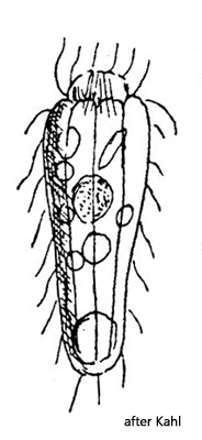
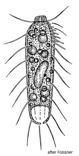
broad oral bulge with rod-shaped extrusomes (3 µm)
oral bulge surrounded by elongated cilia
sparse, long cilia
macronucleus short ellipsoid
contractile vacuole terminal
Rhopalophrya crassa
So far, I have found only two specimens of Rhopalophrya crassa. I found the first specimen in November 2024 in the Simmelried and the second one also in November in the Bussenried. I have no further records to date.
In the little-studied genus Rhopalophrya, Kahl (1926) described small, club-shaped ciliates with a distinct furrowing of the pellicle and long, soft cilia. In all forms, the front end is beak-shaped. Only in the species Rhopalophrya crassa is a distinct oral bulge visible.
The two specimens of Rhopalophrya crassa swam very slowly, with rowing-like movements of the loosely spaced cilia. The somatic cilia are conspicuously long, measuring 13–15 µm. In the specimen from the Bussenried, I could also observe that the cilia around the oral bulge were significantly longer at 20 µm (s. fig. 3). This is also mentioned by Kahl. In both specimens, I could clearly see longitudinal ribs (s. fig. 1 a-f). The extrusomes in the oral bulge of my specimens were 5–6 µm long. The macronucleus was spherical. I could not identify the micronucleus. The contractile vacuole is terminal.
In 2002, Foissner, Agatha, and Berger described an Enchelyodon minutus from Namibia (s. fig. 4), which closely resembles Rhopalophrya crassa in almost all characteristics. However, the authors did not synonymize the species because Enchelyodon minutus has an ellipsoid or kidney-shaped macronucleus, whereas Rhopalophrya crassa has a spherical one. Whether synonymization is justified can only be determined by further findings.
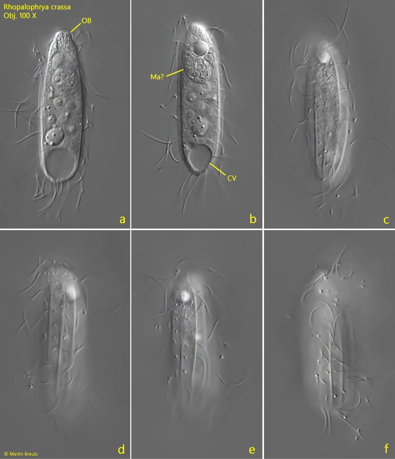
Fig. 1 a-f:Rhopalophrya crassa. L = 43 µm. A different focal planes of a freely swimming specimen. The longitudinal ridges of the body are clearly visible. CV = contractile vacuole, Ma? = probably the macronucleus. Obj. 100 X.
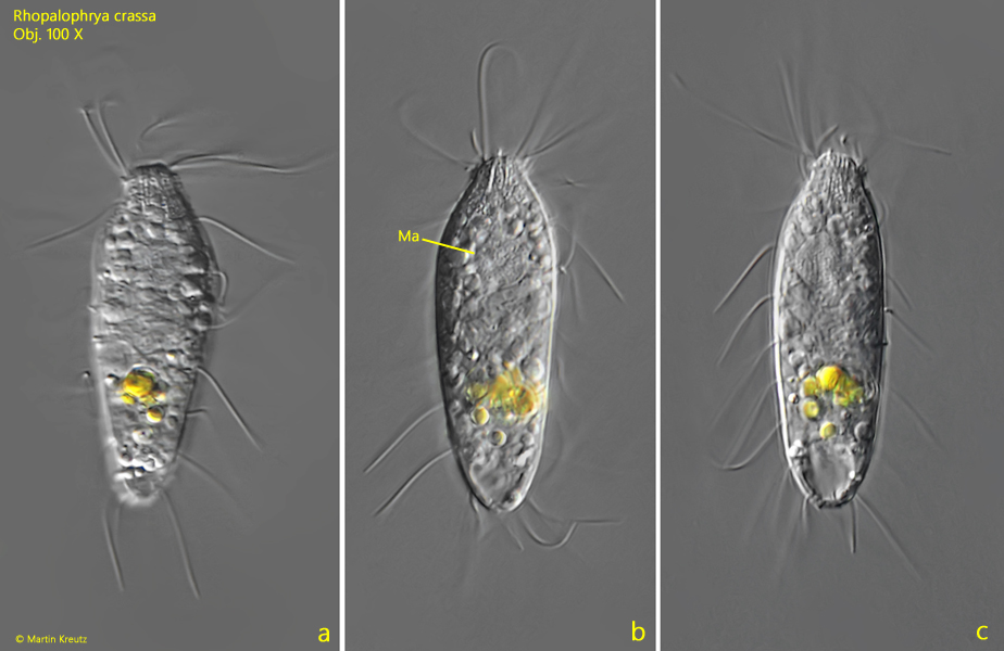
Fig. 2 a-c:Rhopalophrya crassa. L = 43 µm. A second specimen found 2014 in the Bussenried. Ma = macronucleus. Obj. 100 X.
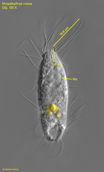
Fig. 3:Rhopalophrya crassa. L = 43 µm. The cilia around the oral bulge are 20 µm long, while the somatic cilia are 13–15 µm long. The extrusomes in the oral bulge are straight rods with a length of 5–6 µm. Ma = macronucleus. Obj. 100 X.
Fig. 4:Enchelyodon minutus, described by Foissner, Agatha and Berger (2002), is likely synonym with Rhopalophrya crassa.