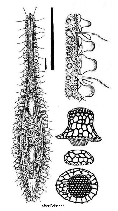
body amphoriform or fusiform, contracted bottle-shaped
dorso-ventrally flattened
oral bulge conical or cylindrical, with apical tip
length 160–230 µm, width 15–40 µm
two widely separated, ellipsoid macronuclei
each macronucleus with one micronucleus
about 12 longitudinal rows of cilia
two types of rod-shaped extrusomes
extrusomes type 1 acicular, 12–16 µm long
extrusomes type 2 thin, straight rods, length 2.5 µm
cells covered by epicortical layer with lepidosomes
two types of complex shaped lepidosomes
dorsal brush three-rowed
contractile vacuole terminal
excretion pore terminal
Sleighophrys pustulata
Sleighophrys pustulata was first described by Foissner from Venezuela in 2005. He found this species in samples from dried-up puddles on a cattle pasture. No other locations for this trachelophyllid ciliate have been known so far.
I found Sleighophrys pustulata in June 2026 in the Gieringer Weiher near Kitzbühel in Austria. This is a largely untouched raised bog. The samples were taken in shallow pools around the pond. In the samples, the specimens accumulated at the surface of the samples.
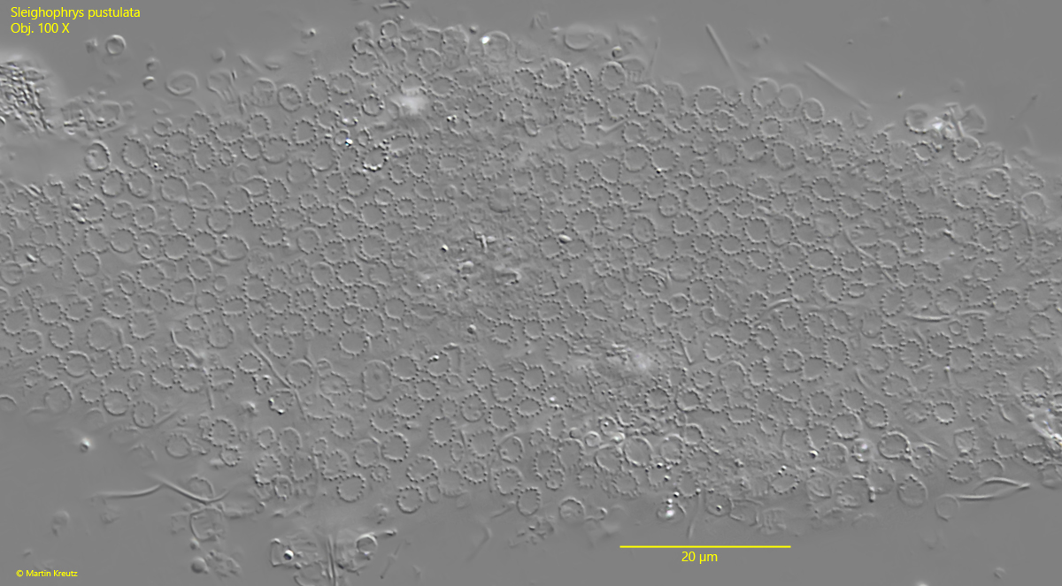
The most striking feature of Sleighophrys pustulata is an approximately 6–8 µm thick epicortical layer that covers the entire body of the ciliate, except for the apical mouth opening. The epicortical layer is a mucous layer into which intricately shaped scales, called lepidosomes, are embedded. In Sleighophrys pustulata, two different types of lepidosomes are present. Their exact shape can only be seen under an electron microscope. The larger form of these lepidosomes is roughly bell-shaped and can also be easily recognized under a light microscope (s. figs. 3, 4 and 5). In top view, they appear as dotted rings, similar to a bracelet (s. fig. 4). This corresponds exactly to the form as described by Foissner.
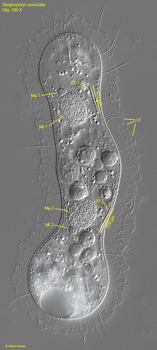
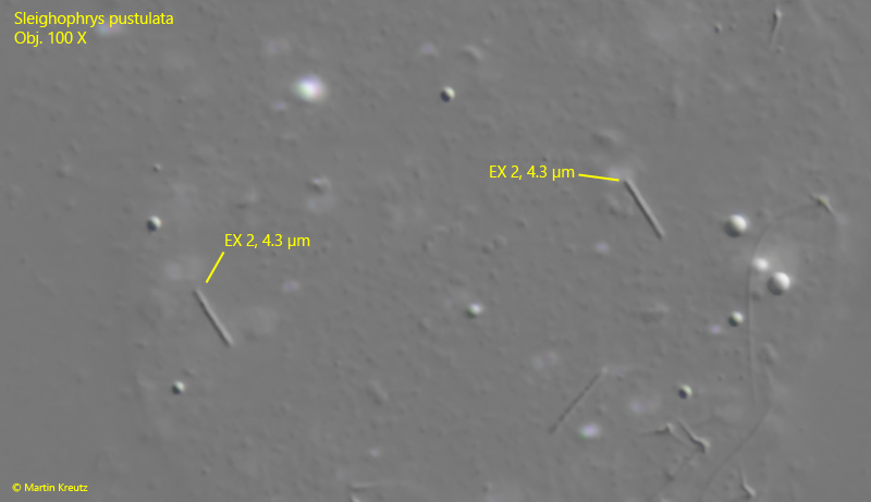
Another important feature is the size and shape of the extrusomes. Foissner observed larger, acicular shaped extrusomes with a length of 12–16 µm and a second form that is rod-shaped and only 2.5 µm long. In my populations, I was also able to observe these forms of extrusomes, but with slightly different lengths. The acicular extrusomes of type 1 were 10–11 µm long (s. fig. 3), while the rod-shaped extrusomes of type 2 were consistently 4.3 µm long (s. fig. 6). Since only one population from Venezuela has been studied so far, I consider these deviations to be the usual variability within the species.
The remaining features also corresponded to the description of Sleighophrys pustulata by Foissner. There are two widely separated macronuclei with an ellipsoidal shape, each with an attached micronucleus (s. fig. 3). The contractile vacuole is terminal (s. fig. 2 a). All the specimens I observed contracted under the coverslip, which caused them to take on a bottle shape and have a correspondingly shortened body length. However, this behavior was also observed by Foissner.
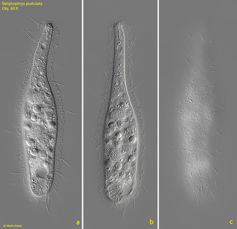
Fig. 1 a-c:Sleighophrys pustulata. L = 130 µm. A slightly contracted, freely swimming specimen. The lepidosomes scattered in the mucilaginous sheath covering the body appear as dotted circles in top view (c). Obj. 60 X.
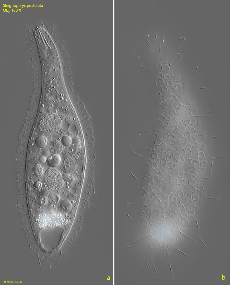
Fig. 2 a-b:Sleighophrys pustulata. L = 130 µm. Two focal planes of the specimen as shown in fig. 1 a-c. Obj. 100 X.
Fig. 3:Sleighophrys pustulata. A squashed second specimen. Two of the acicular shaped extrusomes type 1 (EX 1) are visible with a length of 10.1 µm and 10.9 µm. The nuclear apparatus consists of two separated macronuclei (Ma 1, Ma 2) each with one adjacent micronucleus (Mi 1, Mi 2). LE = lepidosomes. Obj. 100 X.
Fig. 4:Sleighophrys pustulata. A strongly squashed specimen with focal plane on the ring-shaped lepidosomes scattered in the mucilainous layer covering the cell. Obj. 100 X.
Fig. 5:Sleighophrys pustulata. The lepidosomes (LE) with a length of about 4 µm in a strongly squashed specimen. Obj. 100 X.
Fig. 6:Sleighophrys pustulata. The extrusomes of type 2 (EX 2) in a strongly squashed specimen are rod-shaped with a length of 4.3 µm. Obj. 100 X.