Sonderiella 1
Most likely ID: Sonderiella nov. spec.
Synonym: n.a.
Sampling location: Simmelried, Purren pond
Phylogenetic tree: Sonderiella
Diagnosis:
- body ellipsoid, dorso-ventrally flattened
- ventral side flat, dorsal side domed with slight ribs
- length 33–35 µm
- ventral side with 12–15 longitudinal rows of cilia
- dorsal side naked
- mouth opening anterior on ventral side
- oral apparatus consist of two lips
- lower lip sickle-shaped protrudes ventral side
- upper lip U-shaped
- contractile vacuole on right side, above cell equator
- fringe of extrusomes beneath pellicle
- extrusomes straight, rod-shaped, 6.2–6.8 µm long
- macronucleus ellipsoid, posterior on left side
- one spherical micronucleus
- one caudal cilia
No drawings from previous autors available
In May 2021, I found in the Simmelried, in a layer of decomposed leaves, several specimens of a ciliate with the characteristics of the genus Sonderiella. A few months later, in February 2022, I found further specimens in Purren Pond, again among decomposed leaves.
Within the genus Sonderiella, only Sonderiella scandens was described by Kahl in 1928 as a valid species. Kahl found the specimens both in saltwater and in freshwater. The species is only 20–25 µm long and has a broadly oval body shape, as Kahl’s drawing shows (s. fig. 1). So far, there is only a single redescription of Sonderiella scandens available by Jeffrey Silverman from July 2025 (s. Sonderiella scandens, iNaturalist).
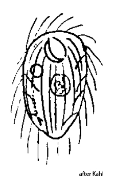
Fig. 1: Drawing of Sonderiella scandens from Kahl, 1928.
On the basis of the description of Sonderiella scandens by Kahl and the video documentation by Jeffrey Silverman, I believe I have found another species of Sonderiella, since its characteristics differ significantly. For the time being I would like to call it Sonderiella 1.
The specimens of my population of Sonderiella were 33–35 µm long and thus about 30% longer than Sonderiella scandens. The body shape is ellipsoid, however with only weakly convex lateral sides, which makes the shape appear slender (s. figs. 2 a-f and 3 a-f). The body is dorso-ventrally flattened. The flat ventral side shows 12–15 longitudinal rows of cilia that run parallel (s. fig. 2 b). The dorsal side is slightly domed with some faintly developed longitudinal ridges.
The most conspicuous feature is the complicated structure of the oral apparatus. It is located apically on the ventral side. It consists of a sickle-shaped lower lip (s. fig. 2 b) that rises clearly above the flat ventral side (s. fig. 3 f). Opposite it is an upper U-shaped lip that is considerably smaller (s. fig. 2 b). Between the lips, a depression leads to the left into the pharynx. The exact function of this arrangement is difficult to discern even in the living specimen. But it seems as if this oral apparatus serves to scrape off bacterial mats. In the process, only the upper U-shaped lip moves against the lower sickle-shaped lip, which is rigid and immobile.
Numerous symbiotic bacteria can be seen in the cytoplasm. They are thick, rod-shaped, and 4–6 µm long (s. fig. 2 f). In some specimens I had the impression that there is another, smaller species of symbiotic bacteria present in the cytoplasm. However, I was not able to document them photographically.
Beneath the pellicle there is a dense fringe of thin, rod-shaped extrusomes (s. figs. 2 c and 2 d). These are 6.2–6.8 µm long and arranged radially. The fringe extends along the entire body margin and is interrupted only by the mouth opening.
The macronucleus is ellipsoid with a length of about 9–10 µm and is always located at the posterior end on the left side (s. fig. 3 d). Attached to it is a round micronucleus. At the posterior end there is an elongated caudal cilium (s. fig. 3 c), which is difficult to see in the living specimen.
Sonderiella 1 differs from Sonderiella scandens mainly in size, the more slender shape, the caudal cilium, and the symbiotic bacteria in the cytoplasm. In addition, the contractile vacuole is located closer to the cell equator than in Sonderiella scandens, where it is situated in the anterior third. I therefore believe that I have found a previously undescribed species, Sonderiella nov. spec. This is also supported by the fact that the species lives in freshwater.

Fig. 2 a-f: Sonderiella 1. L = 35 µm. Different focal planes from ventral of a specimen found in May 2021 in the Simmelried. The ciliation of the ventral side consists of 13 longitudinal rows of cilia (b). The U-shaped upper lip (ULI) and the sickle-shaped lower lip (LLI) of the oral apparatus are visible (b). Beneath the pellicle a fringe of rod-shaped extrusomes with a length of 6.5-6.7 µm is present. CV = contractile vacuole, MO = mouth opening, SB = symbiotic bacteria. Obj. 100 X.
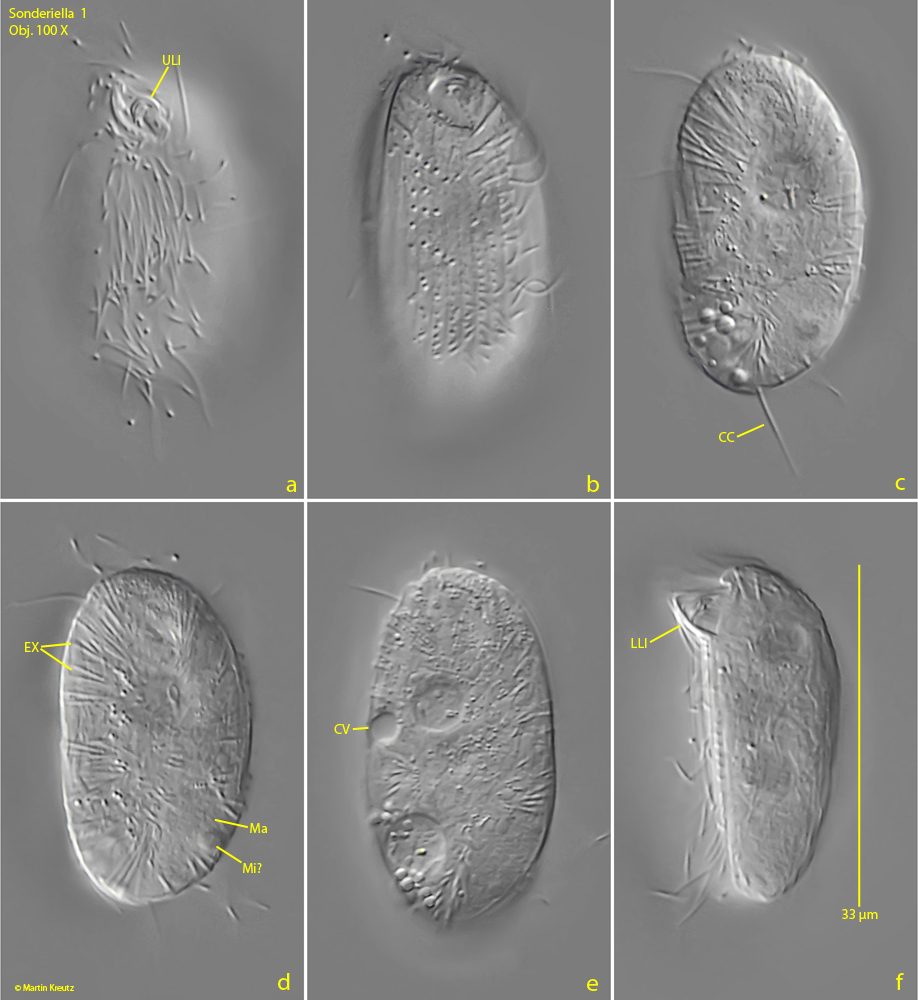
Fig. 3 a-f: Sonderiella 1. L = 33 µm. Different focal planes from ventral of a second specimen found in February 2022 in the Purren pond. Note the caudal cilium (CC) and the protruding lower lip (LLI) of the oral apparatus. CV = contractile vacuole, EX = extrusomes, MO = mouth opening, ULI = upper lip of the oral apparatus. Obj. 100 X