In the Simmelried as well as in my sampling sites Ulmisried and Purren pond, I have found many spathidiid ciliates over the years that I could not identify with the literature available to me. Many of these finds have probably not yet been described. I would like to present these interesting ciliates here as numbered “spathidiid ciliates.” Despite years of observation, I have found only one specimen of some of these spathidiid ciliates.
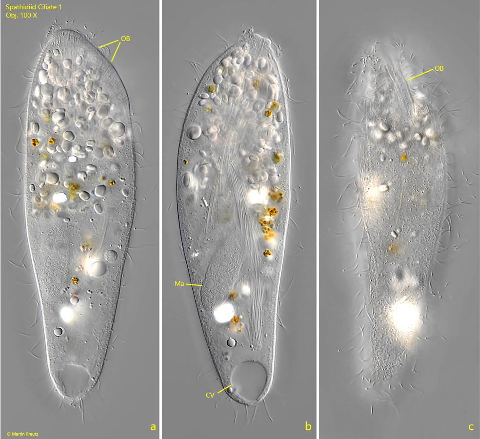
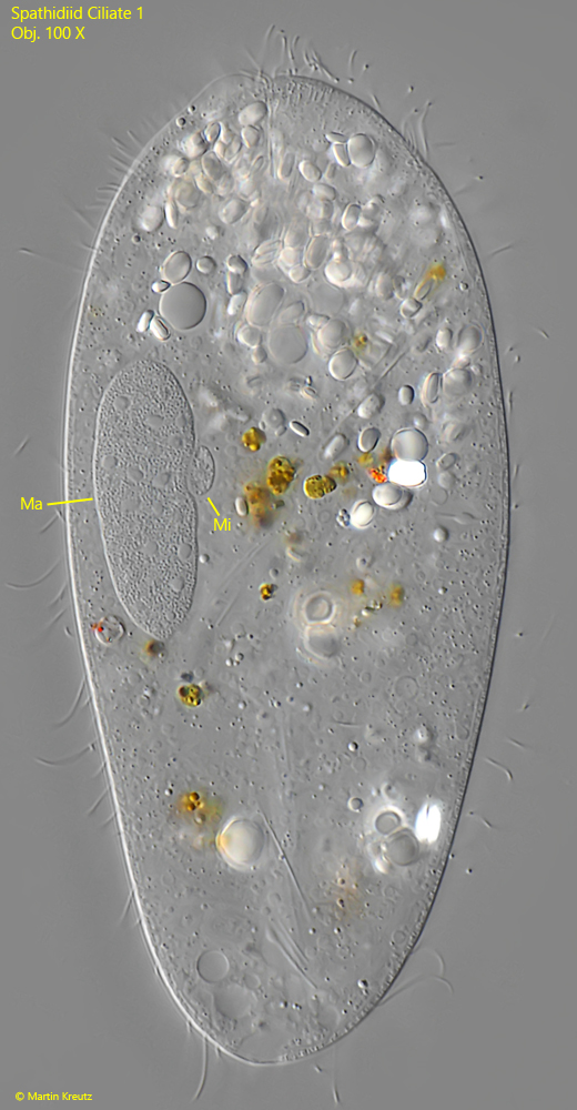
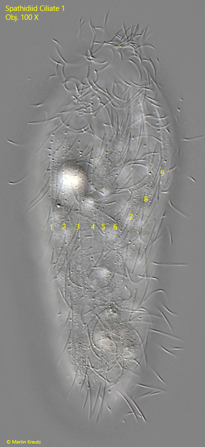
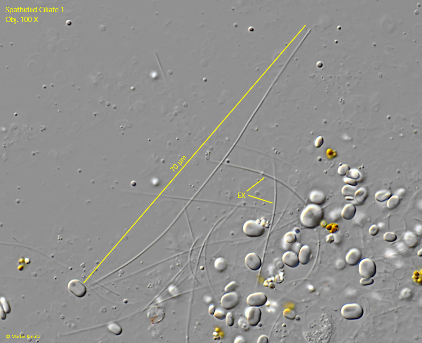
I found spathidiid ciliate 1 in July 2022 in the uppermost mud layer in Simmelried. The specimens were between 95–130 µm long. Freely swimming specimens were slightly flattened in the front third and in the middle of the body. The oral bulge slopes ventrally in an arc (s. fig. 1 a). It bent somewhat to the left side of the body (s. fig. 1 c). I could not clearly identify the dorsal brush. However, I noticed rows of short bristles that are probably part of the dorsal brush. According to my count, there are about 20-24 somatic kineties over the body present (s. fig. 3). Very long and thin extrusomes stand out in the cytoplasm. They are flexible, pliable, and about 70 µm long (s. fig. 4). I could recognize no second type of extrusomes. The macronucleus is elongated ellipsoid. A lens-shaped micronucleus lies adjacent to it (s. fig. 2). The contractile vacuole is terminal with an excretory pore at the posterior pole (s. fig. 1 b).
In the literature, I could not identify any spathidiid species with these characteristics. In particular, the exceptionally long extrusomes of this ciliate stand out. It could therefore be a species not yet described.
Fig. 1 a-c:Spathidiid ciliate 1. L = 126 µm. A freely swimming specimen from right (a), dorsal (b) and from ventral (c). CV = contractile vacuole, Ma = macronucleus, OB = oral bulge. Obj. 100 X.
Fig. 2:Spathidiid ciliate 1. L = 126 µm. The same specimen as shown in fig. 1 a-c slightly squashed. Ma = macronucleus, Mi = micronucleus. Obj. 100 X.
Fig. 3:Spathidiid ciliate 1. L = 96 µm. Focal plane on the longitudinal rows of somatic cilia (1–9) of a second specimen. The total number of rows is about 20-24. Obj. 100 X.
Fig. 4:Spathidiid ciliate 1. The extrusomes (EX) are thin, flexible rods with a length of about 70 µm. Obj. 100 X.