Spathidioides 1

Most likely ID: Spathidioides nov. spec.


Synonym: n.a.


Sampling location: Simmelried


Phylogenetic tree: Spathidiida


Diagnosis: 

  • body sac-shaped, laterally slightly flattened
  • length 110–150 µm
  • Oral bulge with dorsal verucciform elevation
  • macronucleus elongated ellipsoid
  • extrusomes type 1, rod-shaped, 0.40–0.48 µm
  • extrusomes type 2, rod-shaped, 0.20–0.24 µm
  • about 24–30 longitundinal rows of cilia
  • contractile vacuole terminal

No drawings from previous authors available.

In October 2010, I found a specimen of a ciliate among rotting aquatic plants in the Simmelried with the characteristics of the genus Spathidioides. This genus was established by Brodsky (1925) and includes spathidiid ciliates whose oral bulge has on the dorsal side a wart-like elevation. Kahl (1935) described 4 species within this genus:


Spathidioides carinata
Spathidioides sulcata
Spathidioides exsecata
Spathidioides armata


Foissner & Xu (2007) added two more species:


Spathidioides euglenivora
Spathidioides rigida


At the same time, Foissner & Xu note that it is questionable whether separating these species into the genus Spathidioides is necessary, as all the aforementioned species have been only very poorly studied. However, since this has not yet happened, I also classify the ciliate I found in this genus.


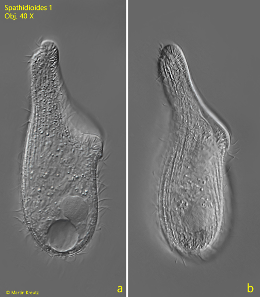
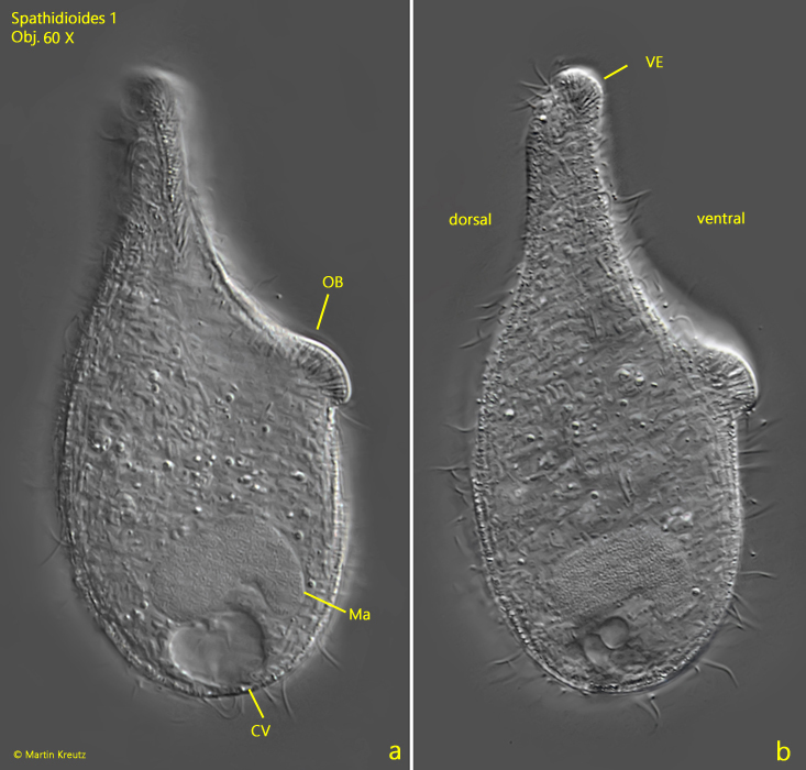
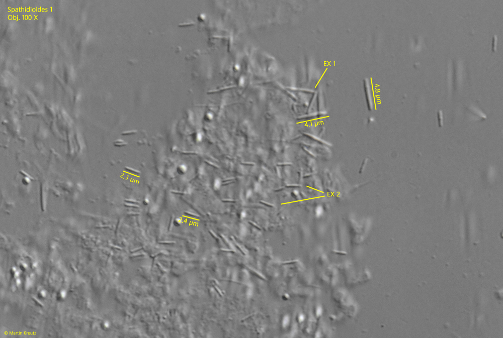
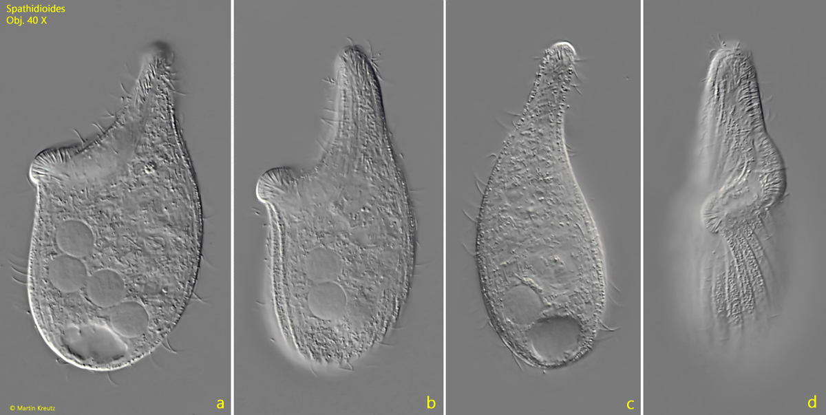
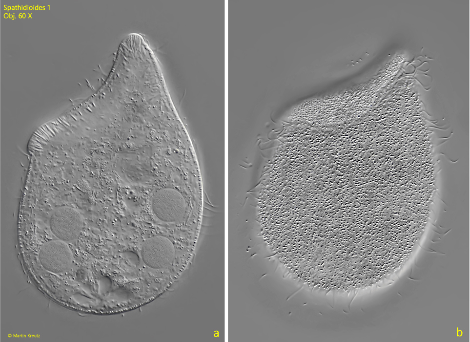
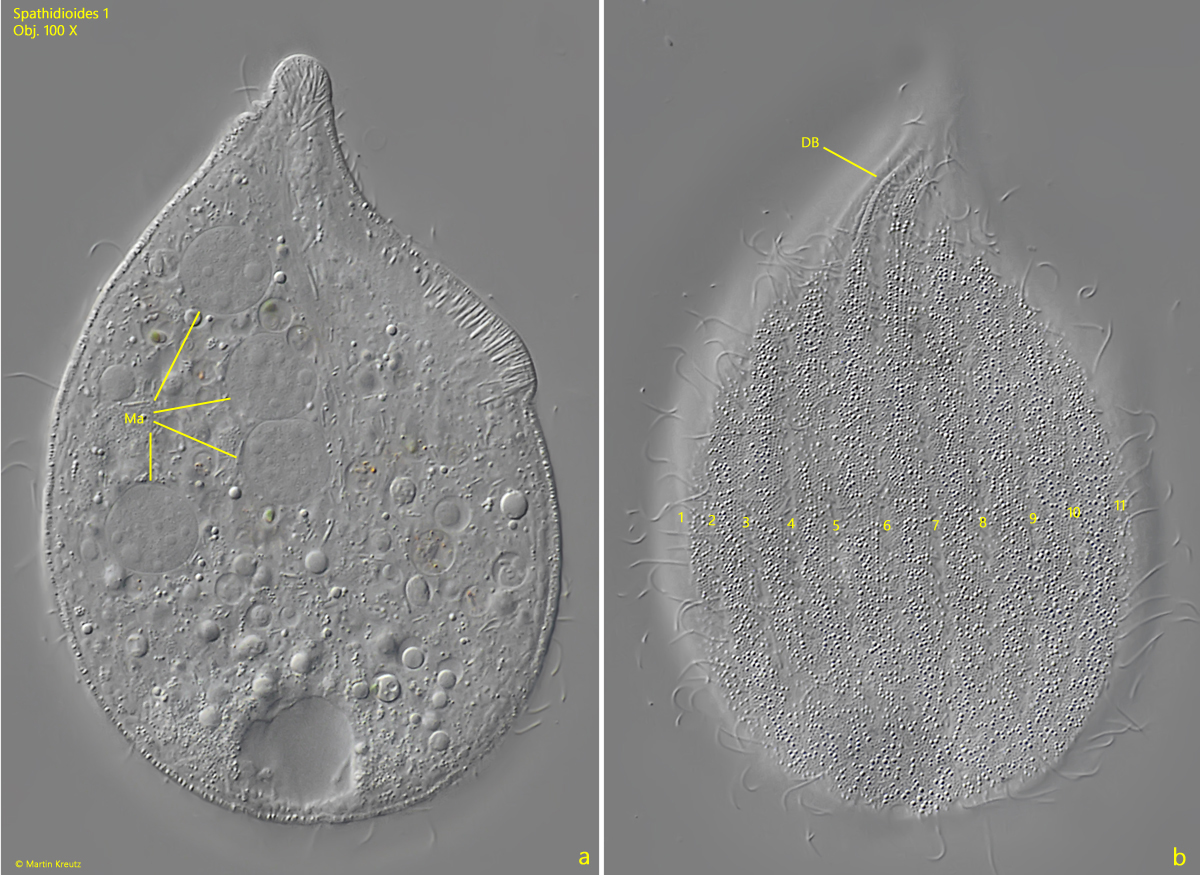
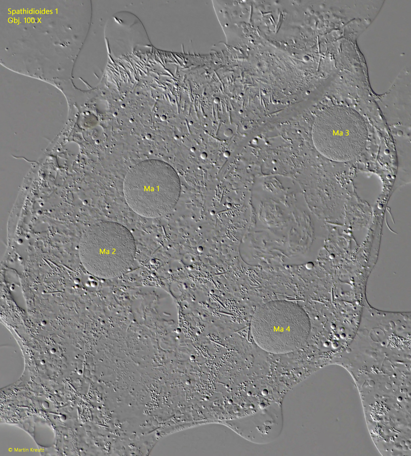
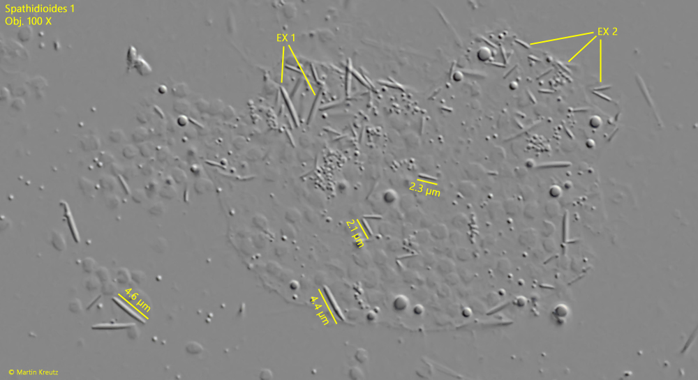
The ciliate from the Simmelried is 145 µm long and has a distinctly curved oral bulge, which rises sharply on the dorsal side and ends there in a wart-like protrusion densely equipped with extrusomes (s. fig. 2 a-b).  The body is sack- or pouch-shaped and somewhat laterally compressed. The macronucleus is elongated ellipsoid and curved (s. fig. 2 a). It is located in the posterior third. The contractile vacuole is terminal. I was able to clearly identify two types of extrusomes (s. fig. 3). Both are rod-shaped. Type 1 is 4.0–4.8 µm long and has slightly tapered ends, while type 2 is only 2.0–2.4 µm long.


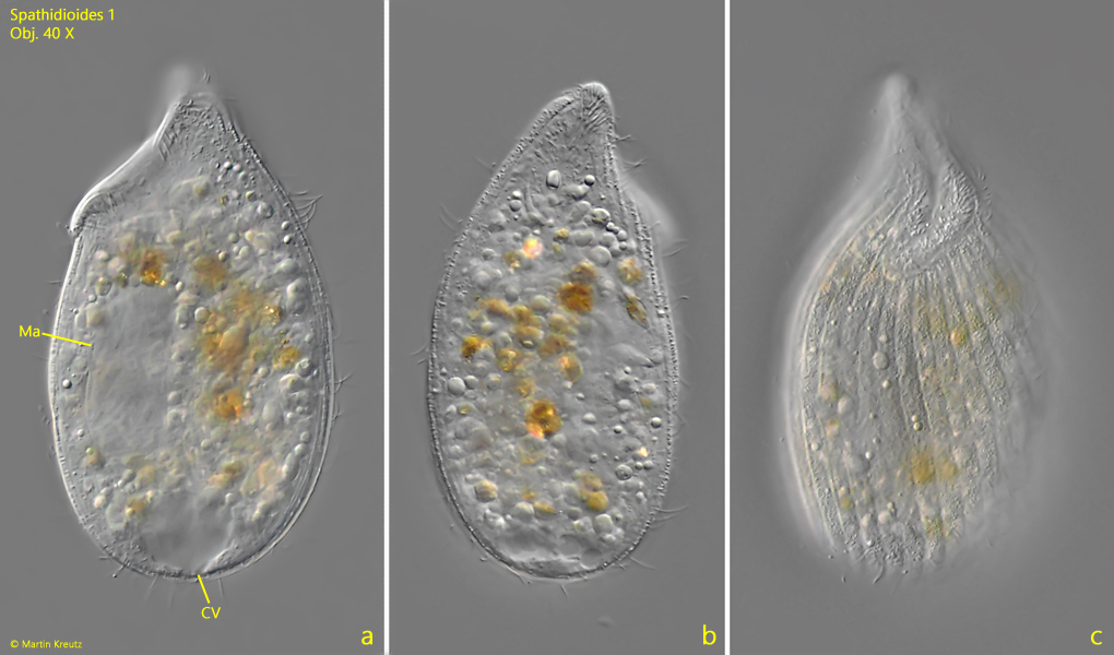
In May 2025, I found a second specimen in the Simmelried with a length of 118 µm (s. fig. 4 a-c). However, I only found this after placing the coverslip, and due to foreign bodies underneath, I could not further reduce the layer thickness. The habitus, macronucleus, and position of the contractile vacuole, however, correspond to the characteristics of the specimen from 2010.


This combination of features does not match any of the previously described species within the genus Spathidioides. Therefore, it must be a previously undescribed, new species, which I provisionally name Spathidioides 1.


A similar species was found by Bruce Taylor in April 2022 near Wakefield, Canada (s. i-Naturalist – Spathidioides). However, he did not provide any information about the size or other characteristics of his find. The similarity to my findings essentially lies in the body shape.

Spathidioides-1

Fig. 1 a-b: Spathidioides 1. L = 145 µm. A freely swimming specimen found in October 2010 in the Simmelried from right. Obj. 40 X.

Spathidioides-1

Fig. 2 a-b: Spathidioides 1. L = 145 µm. The same specimen as shown in fig. 1 a-b at higher magnification. Note the verruciform elevation (EV) at the doral end of the oral bulge (OB). CV = contractile vacuole, Ma = macronucleus. Obj. 60 X.

Spathidioides-1

Fig. 3: Spathidioides 1. The extrusomes type 1 (EX 1) with a length of 4.2–4.8 µm and the extrusumes type 2 (EX 2) with a length of 2.3–2.4 µm in the strongly squashed specimen as shown in fig. 2 a-b. Obj. 60 X.

Spathidioides-1

Fig. 4 a-b: Spathidioides 1. L = 118 µm. The second specimen found in May 2025 in the Simmelried from left (a), dorsal (b) and from ventral (c). CV = contractile vacuole, Ma = macronucleus. Obj. 40 X.

In July 2022, I found a third specimen in the Simmelried (s. fig. 5 a-d). However, this one was significantly smaller at 96 µm in length and had 4 spherical macronuclei (s. figs. 7a and 8). The other characteristics, however, matched those of the two specimens shown above. Therefore, this is very likely a post-conjugant, which was in the reorganization phase of the nuclear apparatus. The fact that this is a third specimen of the same species is particularly evidenced by the extrusomes, which are identical in shape and length to those of the specimens shown above (s. fig. 9).

Spathidioides-1

Fig. 5 a-d: Spathidioides 1. L = 96 µm. A third specimen with four macronuclei from left (a, b), dorsal (c) and from ventral (d). The specimen was found in July 2022 in the Simmelried and is likely a post-conjugant. Obj. 40 X.

Spathidioides-1

Fig. 6 a-b: Spathidioides 1. L = 96 µm. The slightly squashed specimen as shown in fig. 5 a-d from left. Obj. 60 X.

Spathidioides-1

Fig. 7 a-b: Spathidioides 1. The squashed specimen as shown in fig. 5 a-d from right (a) and from dorsal (b). The four globular macronuclei (Ma) are very likely the result of the reorganisation of the nuclear apparatus after a conjugation. On the dorsal side the dorsal brush (DB) is visible as well as 11 longitudinal rows of cilia (1-11). Obj. 100 X.

Spathidioides-1

Fig. 8: Spathidioides 1. In the strongly squashed specimen as shown in fig. 5 a-d the four globular macronuclei are visible (Ma 1-4) but no micronucleus. Obj. 100 X.

Spathidioides-1

Fig. 9: Spathidioides 1. The extrusomes type 1 and type 2 (EX 1, EX 2) in the strongly squashed specimen as shown in fig. 5 a-d. They have the same shape and length as the specimen found in October 2010 (s. fig. 3). Obj. 100 X.