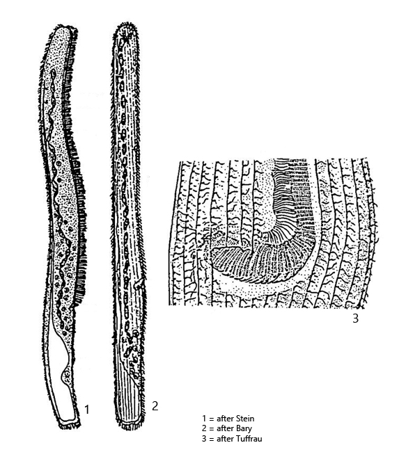
at mouth opening adoral zone bends to right in a hockey stick shape
Spirostomum ambiguum
Spirostomum ambiguum is among the largest ciliates overall, which I encounter at many of my sampling sites. Most of the specimens are found in the sludge zone. In rare cases, I have also observed mass developments.
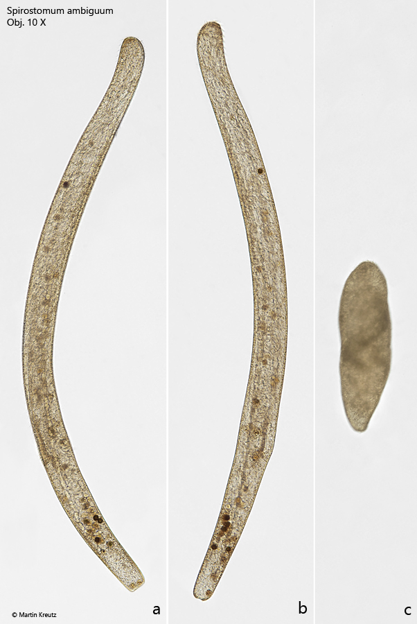
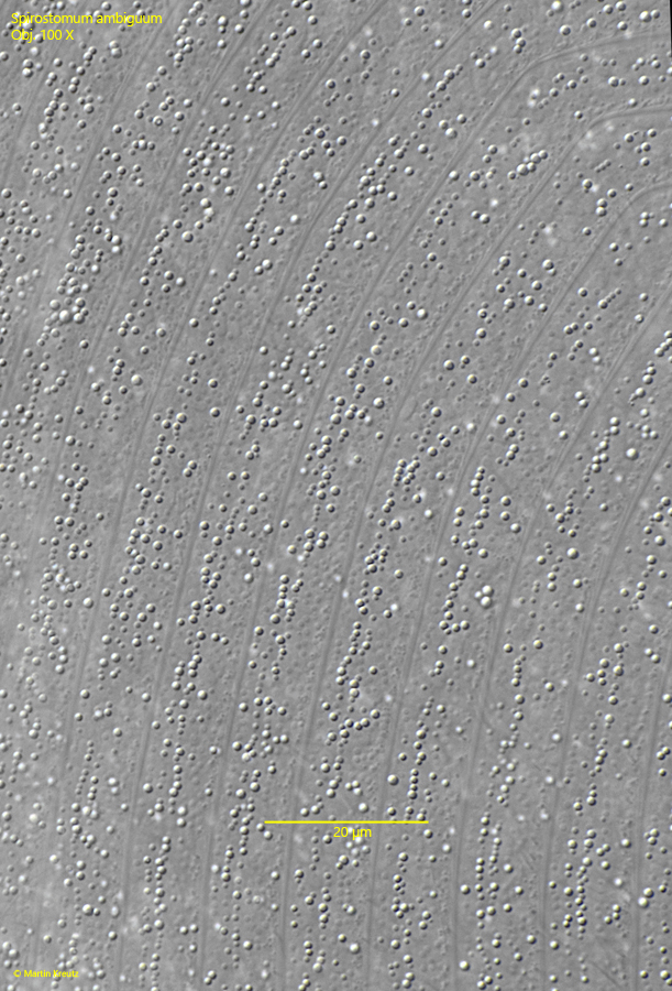
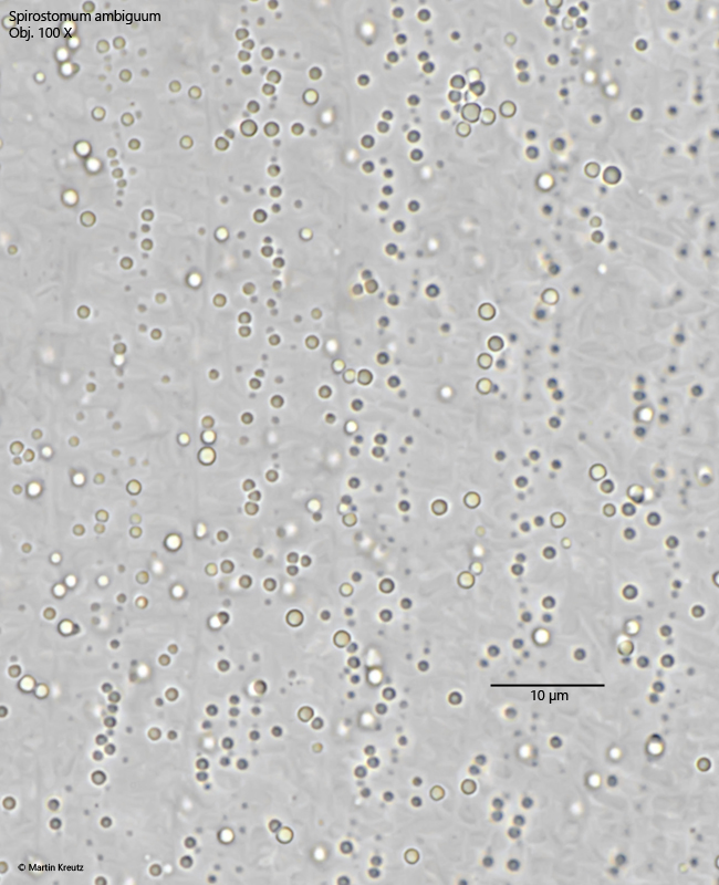
The body of Spirostomum ambiguum is almost parallel-sided along the entire body length, and the posterior end does not taper. Additionally, the specimens are distinctly yellowish-brownish in color (s. figs. 1 a-c and 2). This coloration is caused by the cortical granules, which are yellowish-brownish colored lipid droplets. These lipid droplets, with a maximum size of 2 µm, are arranged in bands between the longitudinal rows of the somatic cilia. Within these bands, the droplets are irregularly arranged (s. figs. 6 and 7).
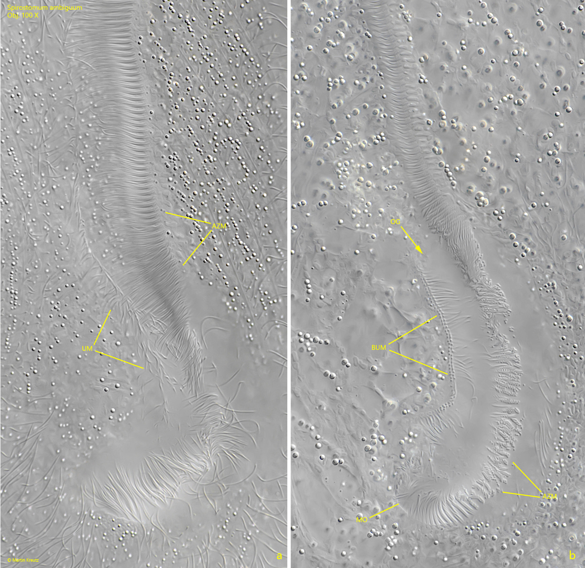
The adoral zone of Spirostomum ambiguum is enormously long and extends from the apical end down to the last third of the body, where the mouth opening is located. The adoral zone lies on the right side of a flat oral groove. Just before the mouth opening, the adoral zone bends sharply to the right and then runs into the cavity of the mouth opening. On the left side of the oral groove is the inconspicuous undulating membrane, which arises from a double row of basal bodies (s. fig. 4 a-b).
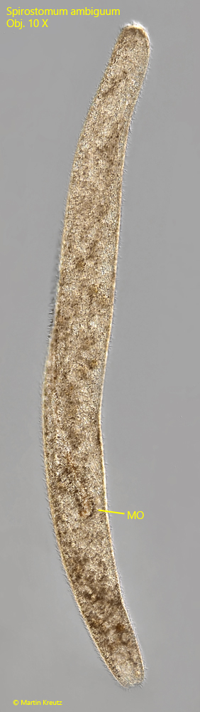
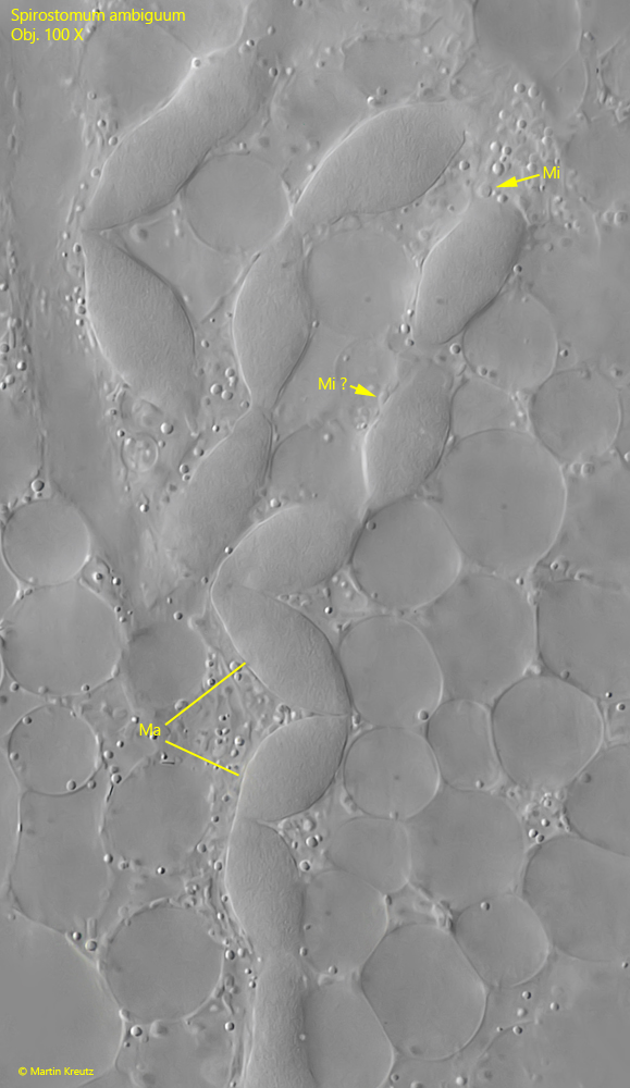
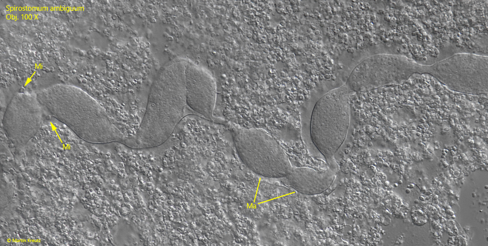
The macronucleus consists of macronuclear nodules that are arranged like a string of pearls. The chain of nodules is about as long as the body. The individual nodes are separated from each other by constrictions, at which the very small micronuclei are usually also found (s. figs. 8 and 9).
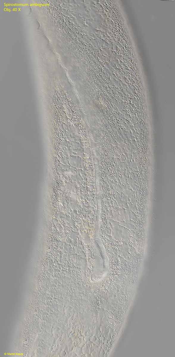
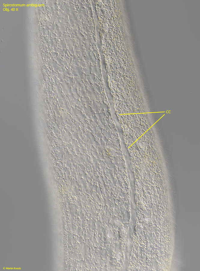
The contractile vacuole is located terminally and has a long collecting canal that extends to the front end of the body. The canal runs on the dorsal side and is easy to see when slightly compressed specimens rotate under the coverslip (s. fig. 5).
Spirostomum ambiguum can be best distinguished from the similar species Spirostomum minus by body length and the length of the adoral zone. Spirostomum minus usually does not exceed 1 mm in length and the adoral zone reaches only 35–50% of the body length.
Fig. 1 a-c:Spirostomum ambiguum. L = 1890 µm. An elongated (a, b) and fully contracted specimen (c) in brightfield illumination. Obj. 10 X.
Fig. 2:Spirostomum ambiguum. L = 1430 µm. A second elongated specimen in DIC. Note the mouth opening (MO) in the posterior third of the body. Obj. 10 X.
Fig. 3:Spirostomum ambiguum. Ventral view on the mouth opening in a slightly squashed specimen. Obj. 40 X.
Fig. 4 a-b:Spirostomum ambiguum. Two focal planes of the mouth opening (MO). The adoral zone of membranelles (AZM) on the right side of the oral groove (OG) bend to the right into the cavity of the mouth opening. on the left side of the oral groove the inconspicuous undulating membrane (UM) is visible. The undulating membrane is formed by a double row of basal bodies (BUM). Obj. 100 X.
Fig. 5:Spirostomum ambiguum. Focal plane on the dorsal collecting canal (CC) of the contractile vacuole. Obj. 40 X.
Fig. 6:Spirostomum ambiguum. In the pellicle, band-shaped longitudinal rows of cortical granules are arranged between the rows of cilia. The granules vary in size (0.1-2 µm) and are irregularly arranged within the bands. Obj. 100 X.
Fig. 7:Spirostomum ambiguum. The cortical granules in a second specimen in brightfiled illumination. Note the slight yellowish-brownish coloration of the granules. Obj. 100 X.
Fig. 8:Spirostomum ambiguum. The macronuclear nodules (Ma) in a squashes specimen. The micronuclei (Mi) are small and hard to see. Obj. 100 X.
Fig. 9:Spirostomum ambiguum. The macronuclear nodules (Ma) and micronuclei (Mi) of a second specimen. Obj. 100 X.