Spirotaenia turfosa

West & G.S. West, 1898

Most likely ID: n.a.


Synonym: n.a.


Sampling location: Schwemm (Austria)


Phylogenetic tree: Spirotaenia turfosa


Diagnosis: 

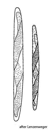
  • cells cylindrical or bread-shaped
  • cells very slightly curved, apices rounded
  • cells 9–13 times as long as wide
  • length 62–100 µm
  • chloroplast with 1.5–2 turns
  • chloroplast with several pyrenoids
  • nucleus central
Spirotaenia-turfosa
Spirotaenia turfosa

In a 4‑week‑old sample from the Schwemm Moor in Austria, I found two dividing stages of Spirotaenia turfosa in June 2026. So far, I have not been able to find any further specimens.


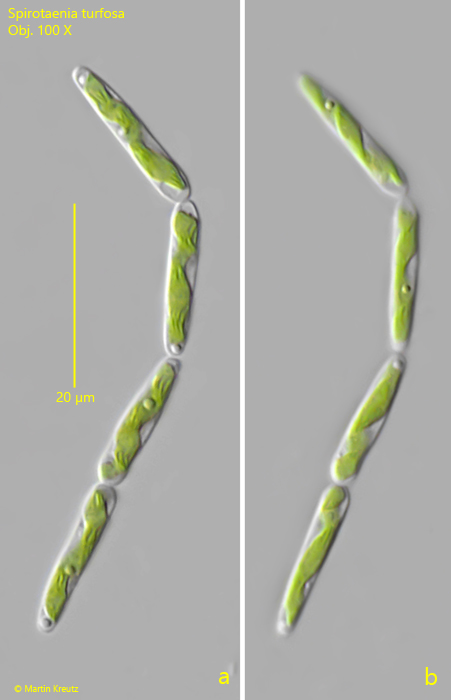
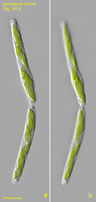
The dividing cells were 15–28 µm long. This is significantly smaller than the range of 62–100 µm given by Lenzenweger (2003), but understandable for dividing cells. In the cells, the spiraling chloroplast is clearly recognizable. In the small cells (s fig. 1 a-b) there were 1.5–2 turns, while in the larger cells (s. fig. 2 a-b) I was able to discern 2–3 turns. In the larger cells this is one more than indicated by Lenzenweger. The length-to-width ratio in all dividing cells is 9. I was unable to identify pyrenoids in the spirals of the chloroplasts.


As alternative species, only Spirotaenia minuta and Spirotaenia parvula come into consideration. Spirotaenia minuta reaches a length of 15–25 µm and shows 2–3 turns. However, two cells are always slightly offset from one another within a common mucilage envelope. This was not the case with my specimens, which is why I excluded this species.

 
In Spirotaenia parvula the chloroplast shows only 1–1.5 turns and the cells are 15–30 µm long. The length-to-width ratio in this species is 5–7. Since I was able to discern more turns of the chloroplast in my specimens and because the length-to-width ratio is 9, I have likewise ruled out Spirotaenia parvula.

Due to a lack of alternatives, identification as Spirotaenia turfosa is the most likely, even though there are some deviations from the description by Lenzenweger, who ultimately reproduces the data from the original description of Spirotaenia turfosa by West & West from the year 1898.
Spirotaenia-turfosa

Fig. 1 a-b: Spirotaenia turfosa. L = 15–18 µm. Two focal planes of 4 specimens in the process of cell division. The chloroplasts in the cells are spiralized with 1.5–2 turns. Obj. 100 X.

Spirotaenia-turfosa

Fig. 2 a-b: Spirotaenia turfosa. L = 27–30 µm. Two focal planes of 2 specimens in the process of cell division. The chloroplasts in the cells are spiralized with 2–3 turns. Obj. 100 X.