I find Spongomonas discus exclusively as growth on the floating coverslip. Depending on how long the coverslip has floated, the growth can become very dense.
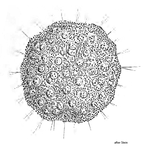
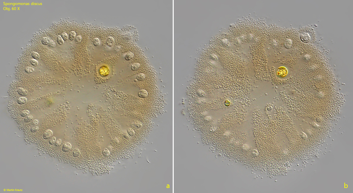
Stein’s description of Spongomonas discus is extremely meager. Essentially, he refers to the round, disc-shaped growth form and to his illustration (s. above). According to my observations, the colonies have a diameter of 50–200 µm depending on age. In my population the colonies were always colored brown or yellow-brown. The mass in which the flagellates are embedded consists of countless small spheres with a diameter of about 1 µm. The flagellates are always located close to the surface of the colonies. Colonies that have grown on the floating coverslip are viewed, so to speak, from below. However, when focusing through, one can see that the colonies are dome-shaped.
According to my observations, the flagellates themselves are either almost spherical when they contract, or long ellipsoid in the extended state. According to my observations, they possess two contractile vacuoles. The first is located at the posterior end and the second in the middle of the body (s. fig. 4). The flagellates are about 9–12 µm long when extended. The equally long flagella measure about 25 µm. The base of the flagella is enclosed by a collar of cytoplasm (s. fig. 5).
Stein (1878) described the species Spongomonas intestinum (now Phalansterium intestinum) Spongomonas uvella and Spongomonas discus. He distinguishes these species according to their growth form. Both Spongomonas intestinum (= Phalansterium intestinum) and Spongomonas uvella form finger-shaped extensions at the ends of which the flagellates sit. In this case, the base of the colonies is stalk-shaped. I was never able to observe this appearance in my population of Spongomonas. Instead, the colonies lay against the coverslip with their entire circular surface, and there was no formation of finger- or grape-shaped extensions, not even in older colonies. Therefore, I believe that this is Spongomonas discus according to Stein’s description.
Fig. 1 a-b:Spongomonas discus. D = 130 µm (of colony). Two focal planes of a colony. Obj. 60 X.
Fig. 2:Spongomonas discus. D = 130 µm (of colony). The same colony a shown in fig. 1 a-b at higher magnification. The flagellates are located near the margin of the colony. Obj. 100 X.
Fig. 3:Spongomonas discus. D = 140 µm (of colony). A second colony with many active flagellates, which have stretched out their flagella (F). Obj. 100 X.
Fig. 4:Spongomonas discus. A section of the colony as shown in fig. 3. The flagellates are about 11 µm long, have 2 contractile vacuoles (CV 1, CV 2) and two flagella (F1, F2) of equal length. Obj. 100 X.
Fig. 5:Spongomonas discus. A flagellate with a collar (CO) surrounding the base of the flagella. Obj. 100 X.