Staurastrum anatinum is an extremely variable species, as can be seen from the numerous described varieties and forms. However, since it is known that the size and shape of Staurastrum anatinum adapt to the respective habitat, it remains unclear to what extent the division into varieties is justified.
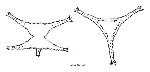
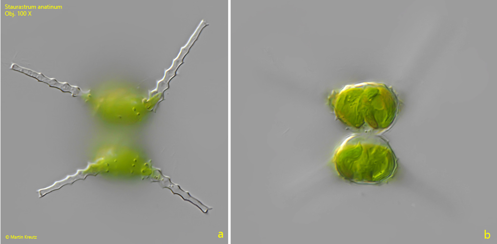
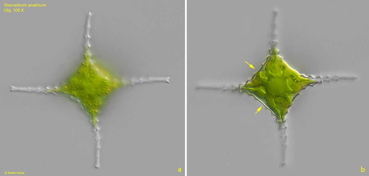
Staurastrum anatinum can easily be confused with Staurastrum pingue and Staurastrum planctonicum, which also have a planktonic lifestyle. An important characteristic of Staurastrum anatinum, however, is the shape of the semi-cells in lateral view. They appear trapezoidal or almost spherical (s. fig. 1 b), but by no means bell-shaped, as is the case with Staurastrum pingue and Staurastrum planctonicum. In apical view, it can also be seen that the sides between the arms in Staurastrum anatinum are almost straight or at most slightly concave (s. fig. 2 b). In the other two species, these sides are distinctly concave.
Fig. 1 a-b:Staurastrum anatinum. L = 53 µm (with arms). Lateral view on the arms (a) and on the almost spherical shape of the semi-cells (b). Obj. 100 X.
Fig. 2 a-b:Staurastrum anatinum. W = 61 µm (with arms). Apical view of the specimen as shown in fig. 1 a-b. The specimen is 4-radiate. Note the almost straight sides between the arms (arrows). Obj. 100 X.