numerous, parietal chromatophores, golden brown, orange or olive
often a red or orange spherical accumulation present
nucleus central, often with visible chromosomes
Stylodinium globosum
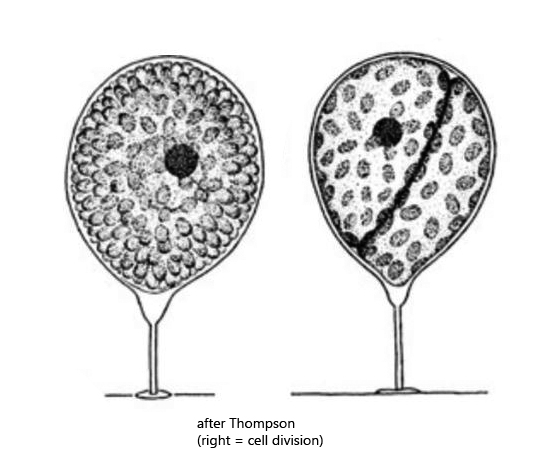
Stylodinium globosum is a sessile dinoflagellate, which settles with a stalk on solid substrates. Mostly these are algal filaments. When screening samples at low magnification, Stylodinium can easily be mistaken for a cyst of other (mobile) dinoflagellates, which can also be spherical. However, the important distinguishing feature is the stalk. The species seems to be very rare. I have found it only once in June 2022 in the Simmelried. I cannot rule out the possibility that I had missed it earlier. Stylodinium globosum is propagated by mobile swarmers, which are released after a preceding cell division. The swarmers are supposed to have the typical shape of dinoflagellates. However, I could neither observe cell division nor the swarmers.
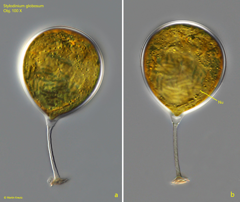
Fig. 1 a-b:Stylodinium globosum. D = 32 µm. Two focal planes of a specimen detached from the substrate. Nu = nucleus with condensed chromosomes. Obj. 100 X.
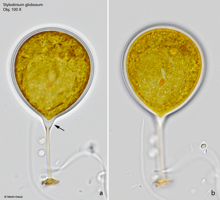
Fig. 2 a-b:Stylodinium globosum. D = 31 µm. Two focal planes of a second specimen in brightfield illumination. Note the characteristic thickening at the junction between stalk and cell (arrow). Obj. 100 X.
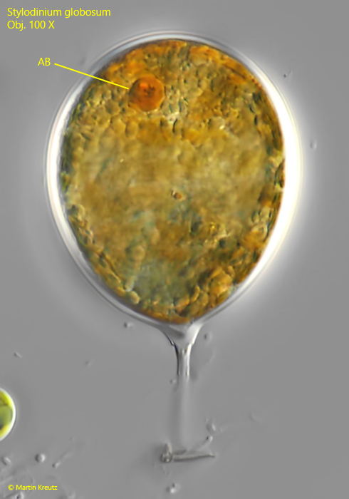
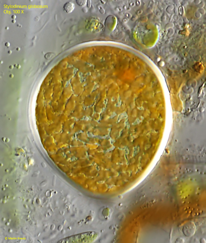
Fig. 3:Stylodinium globosum. D = 35 µm. In many specimens an accumulation body (AB) was present colored red or orange. Obj. 100 X.
Fig. 4:Stylodinium globosum. D = 34 µm. Focal plane on the parietal chromatophores of a slightly squashed specimen. Obj. 100 X.