Tetrahymena pyriformis is one of the best-studied ciliates overall, as this species is cultured in many laboratories around the world for genetic, morphological, and biochemical studies. This model organism is easy to propagate and undemanding in cultures. However, in the “wild,” I have only found Tetrahymena pyriformis twice. The images shown below come from a mass development of Tetrahymena pyriformis in a petri dish with cut stems of a water lily. The specimens fed both on the bacteria forming and on the decomposing plant matter.
The specimens of my population were 40–60 µm long and ovoid in shape. Foissner et al. (1994) report that specimens in culture can also grow up to 90 µm long. Tetrahymena pyriformis can utilize a wide variety of food sources. Besides bacteria, this ciliate also eats flagellates or parasitizes fish and amphibians. There, the ciliate feeds on mucus, blood, serum, and tissue.
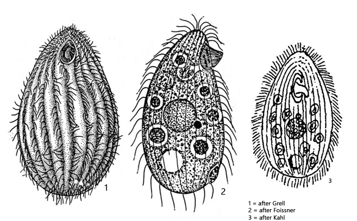
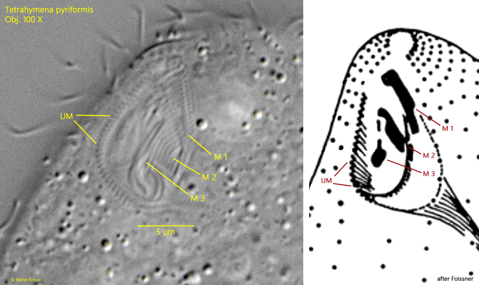
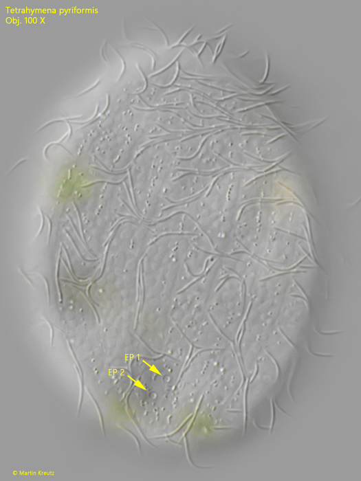
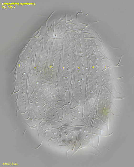
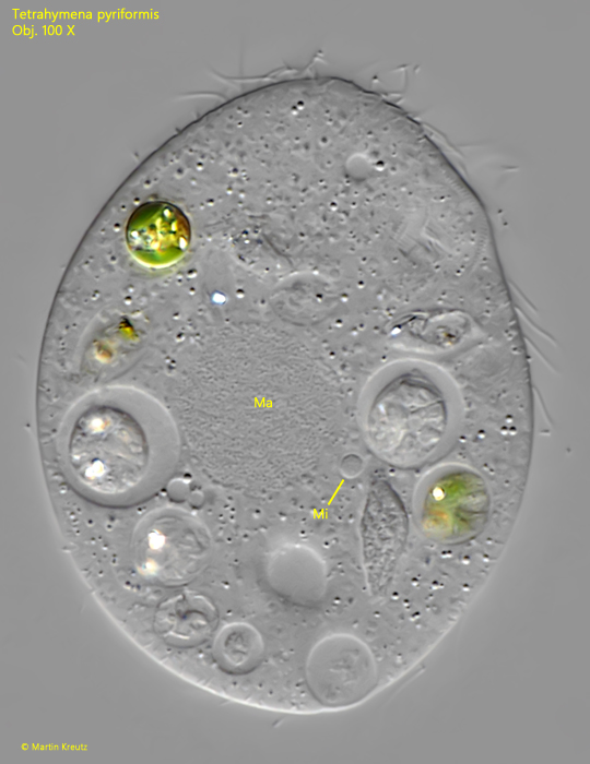
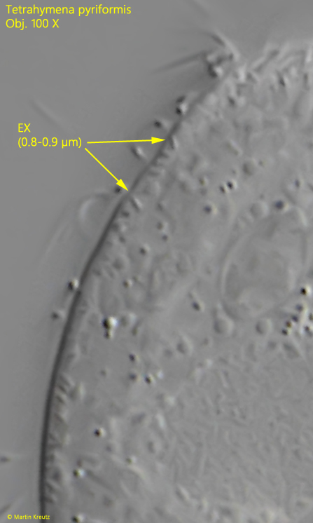
For identification of Tetrahymena pyriformis, the structure of the oral apparatus, the nuclear apparatus, and the number of kineties (longitudinal rows of cilia) are important. The oral opening in Tetrahymena pyriformis is quite small, and it must be examined in squashed specimens at high magnification to recognize the details. It consists of three adoral membranelles of different lengths and is enclosed on the right side by an undulating membrane (s. figs. 3 a and 5). According to Foissner et al., the number of kineties should be between 10-25. I was able to identify 7 kineties on the dorsal side (s. fig. 7), which suggests 14–18 kineties for the entire body. The macronucleus is centrally located with an attached micronucleus, which is sometimes difficult to identify (s. fig. 8). The contractile vacuole is subterminal on the dorsal side (s. fig. 4 a-b) and always had two excretory pores in my population (s. fig. 6). The extrusomes under the pellicle are inconspicuous, rod-shaped, and according to my measurements only 0.8-0.9 µm long (s. fig. 9).
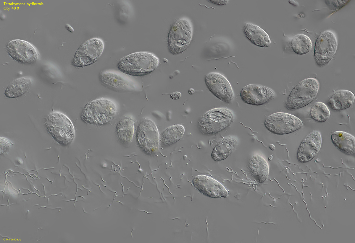
Fig. 1:Tetrahymena pyriformis. L = 44–62 µm. An agglomeration of specimens is feeding on a mass development of spirilla. Obj. 40 X.
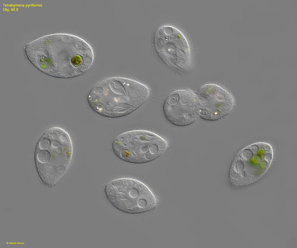
Fig. 2:Tetrahymena pyriformis. L = 49–65 µm. Some freely swimming specimens. One specimen is in the process of cell division. Obj. 60 X.
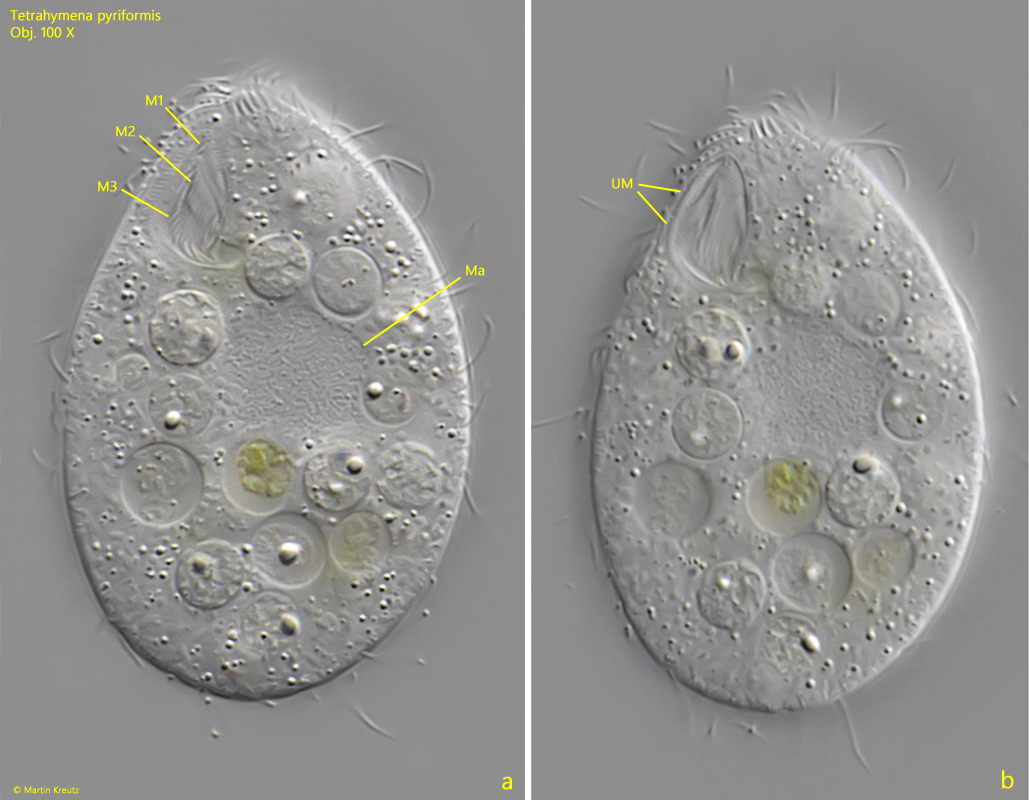
Fig. 3 a-b:Tetrahymena pyriformis. L = 58 µm. Two focal planes of a slightly squashed specimen from ventral. The oral apparatus is consisting of the 3 adoral membranelles (M 1 –M 3) and an undulation membrane (UM) on the right side. Ma = macronucleus. Obj. 40 X.
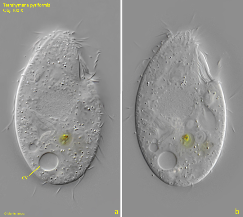
Fig. 4 a-b:Tetrahymena pyriformis. L = 51 µm. Two focal planes of a slightly squashed specimen from right. The contractile vacuole (CV) is located sub-terminal dorsally. Obj. 100 X.
Fig. 5:Tetrahymena pyriformis. The oral apparatus in detail. The arrangement of three adoral membranelles (M 1–M 3) and the undulating membrane (UM) in comparison to a schematic drawing. Obj. 100 X.
Fig. 6:Tetrahymena pyriformis. Focal plane on the two excretion pores (EP 1, EP 2) of the contractile vacuole on the dorsal side. Obj. 100 X.
Fig. 7:Tetrahymena pyriformis. The dorsal side of a slightly squashed specimen with 7 longitudinal rows of cilia (1–7). Obj. 100 X.
Fig. 8:Tetrahymena pyriformis. The spherical macronucleus (Ma) and the adjacent micronucleus (Mi) in a squashed specimen. Obj. 100 X.
Fig. 9:Tetrahymena pyriformis. The inconspicuous extrusomes (EX) are rod-shaped with a length of 0.8–0.9 µm. Obj. 100 X.