Tetrasiphon hydrocora is described as a rare species. I was able to find Tetrasiphon hydrocora only once in June 2025 in the Schwemm, a protected bog area in Austria.
Tetrasiphon hydrocora is restricted to sites with a rich flora of desmids, as the species mainly feeds on these algae. Large Micrasterias species can also be phagocytized as a whole.
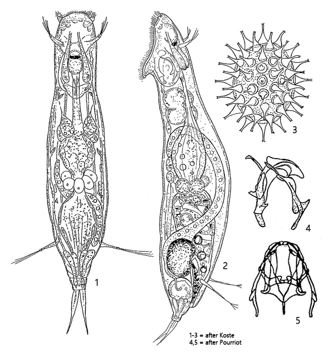
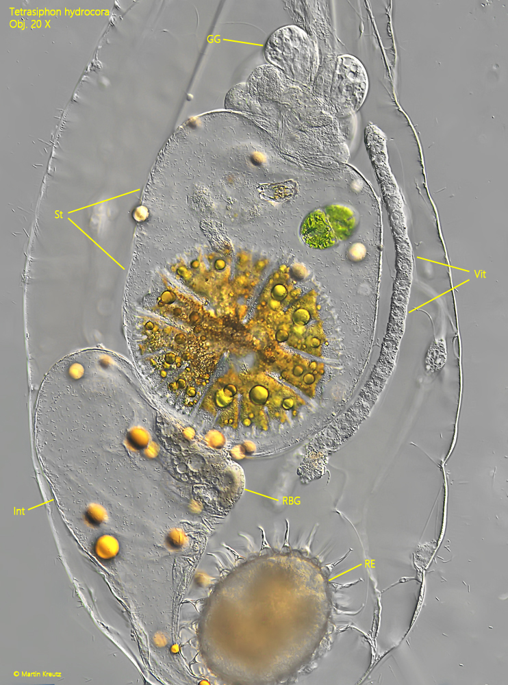
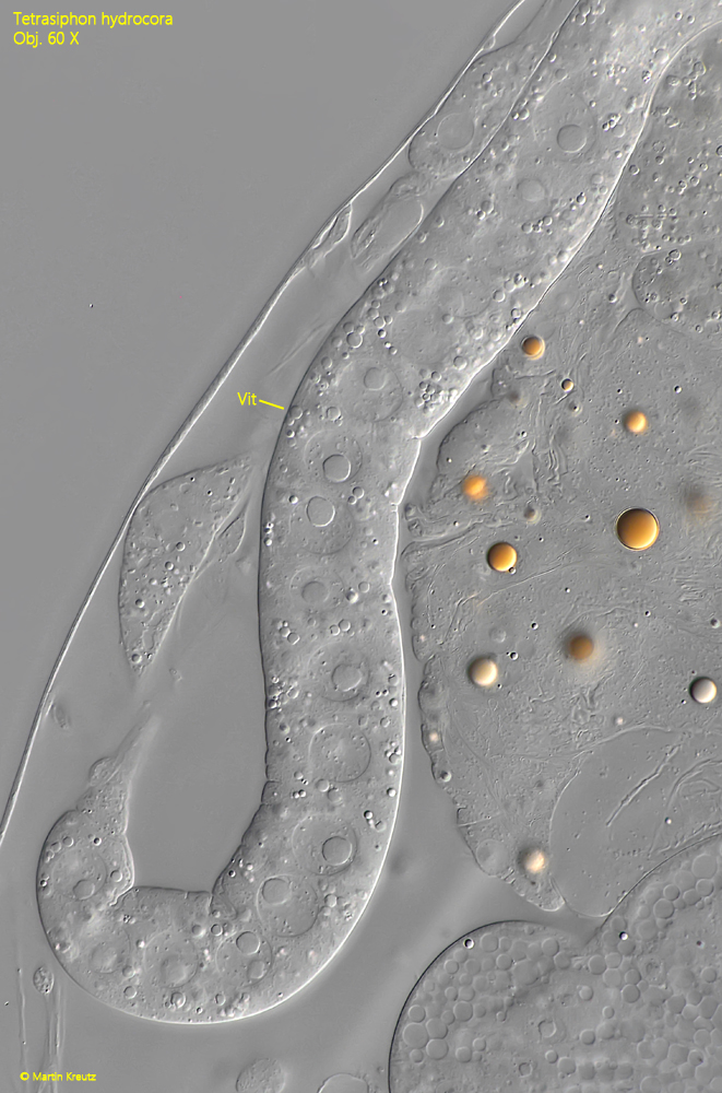
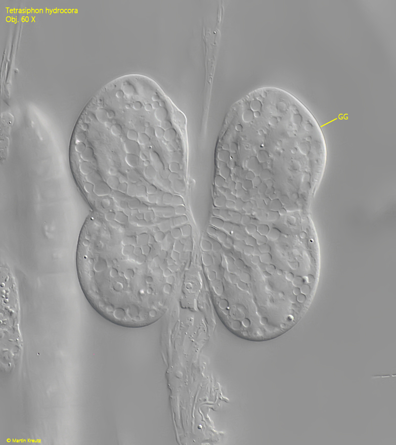
Tetrasiphon hydrocora is very large, up to 1 mm in length, and easy to find in the samples. The specimens also crawl or burrow into slimy detritus flakes. The body has a soft cuticle and is very flexible. The specimens move by crawling. I could not observe any free swimming specimens. The lumen in the body appeared slightly yellowish. The long, tubular vitellarium, which takes up about 60-70 % of the body length, is striking (s. figs. 3 and 7). The stomach is large and sac-shaped. In my specimens it contained exclusively desmids. There were often several orange drops of oil on the outer stomach wall (s. fig. 3). The gastric glands are kidney- oder dumbbell-shaped. They lie close together, so that they appear as a pair in the shape of a butterfly (s. fig. 8).
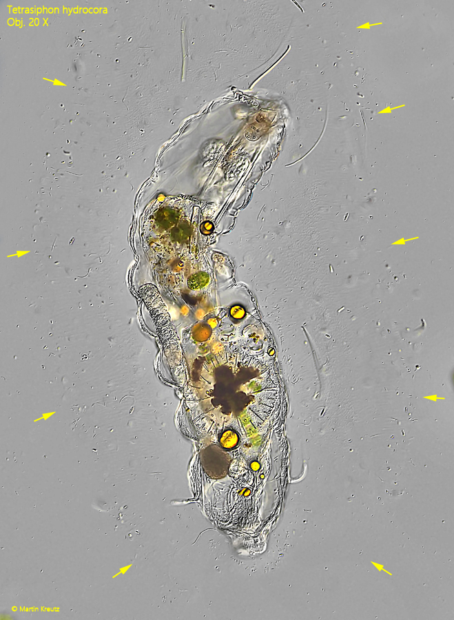
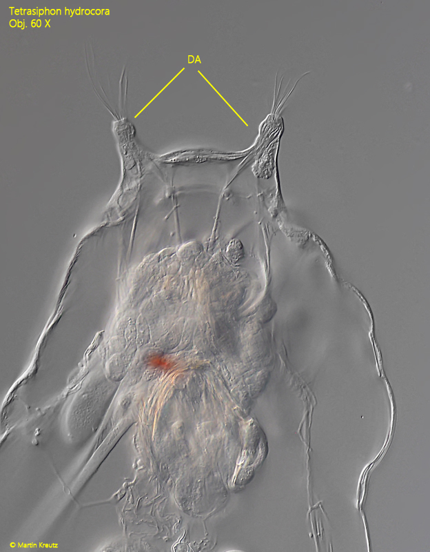
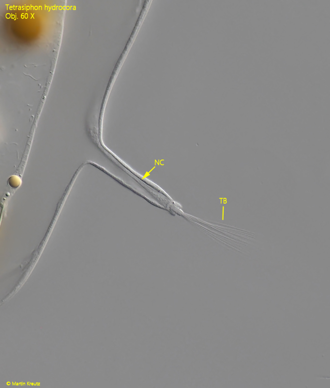
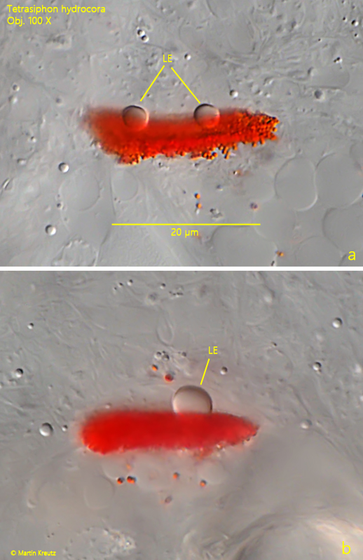
Another striking feature of Tetrasiphon hydrocora are the strongly elongated dorsal and lateral antennae. The tactile bristles sit on elongated stalks, in the case of the lateral antennae these are even tentacle-like (s. figs. 5 and 6). There is a cerebral eyespot with a lens, which is plate-shaped. I have found specimens with one lens but also with two lenses (s. fig. 12 a-b). In my population the specimens with two lenses dominated. The body is surrounded by a thick mucous sheath, which appeared finely granular in the DIC (s. fig. 2). Its dimensions can only be recognized by the adhering bacteria.
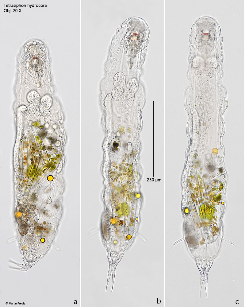
Fig. 1 a-c:Tetrasiphon hydrocora. L = 960 µm. A freely moving specimen from ventral. Obj. 20 X.
Fig. 2:Tetrasiphon hydrocora. The gelatinous sheath of this specimen is covered with bacteria. The approximate outline of the sheath is indicated by arrows. Obj. 20 X.
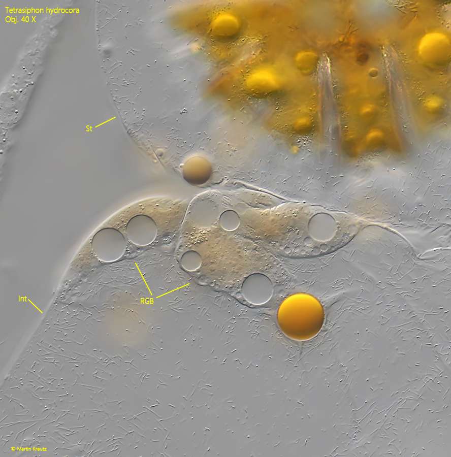
Fig. 3:Tetrasiphon hydrocora. At the narrow transition between the stomach (St) and the intestine (Int) is a ring of glands located that are brownish in color (RGB). GG = gastric glands, RE = resting egg, Vit = vitellarium. Obj. 20 X.
Fig. 4:Tetrasiphon hydrocora. The ring of brownish colored glands (RGB) located at the transition between the stomach (St) and the intestine (Int) in detail. Obj. 40 X.
Fig. 5:Tetrasiphon hydrocora. The dorsal antennae are elongated and located on the dorsal side of the head. Obj. 60 X.
Fig. 6:Tetrasiphon hydrocora. The lateral antennae are tentacle-like elongated. Note the nerve cell (NC) in the antenna and the tactile bristles (TB). Obj. 60 X.
Fig. 7:Tetrasiphon hydrocora. The ribbon-shaped vitellarium (Vit) with large nuclei. Obj. 60 X.
Fig. 8:Tetrasiphon hydrocora. The gastric glands (GG) of this specimen are dumbbell-shaped and the pair of them are butterfly-shaped. Obj. 60 X.
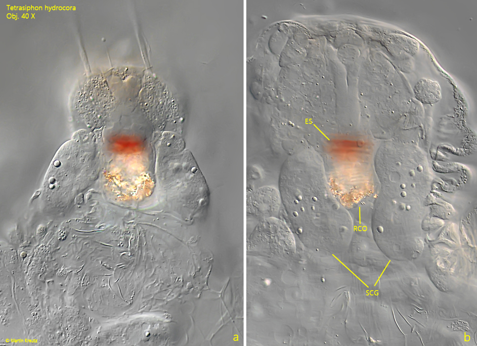
Fig. 9 a-b:Tetrasiphon hydrocora. Below the eyespot (ES) the pair of sub-cerebral glands (SCG) are visible and the retrocerebral organ (RCO). Obj. 40 X.
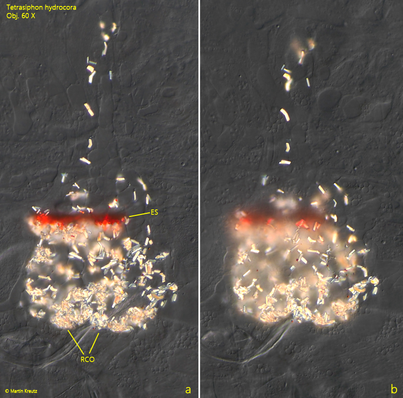
Fig. 10 a-b:Tetrasiphon hydrocora. Two focal planes of the retrocerebral organ (RCO) below the eyespot (ES). The retrocerebral organ is is filled with birefringent crystals, which light up brightly in the DIC. Obj. 60 X.
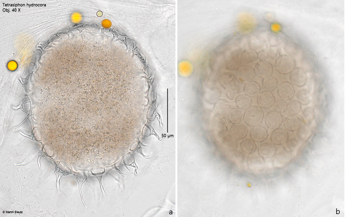
Fig. 11 a-b:Tetrasiphon hydrocora. Two focal planes of a resting egg (RE). The eggs are covered with spines, which have a circular base (b). The spines end in a very fine double point at the distal end. Obj. 40 X.
Fig. 12 a-b:Tetrasiphon hydrocora. The eyespot can have two (a) or only one (b) lenses (LE). Obj. 100 X.
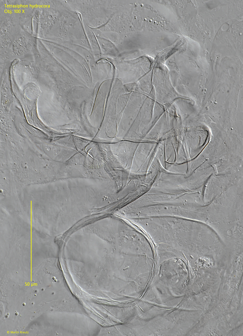
Fig. 13:Tetrasiphon hydrocora. The complex structure of the trophi in a strongly squashed specimen. Obj. 100 X.