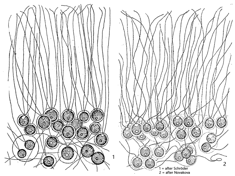
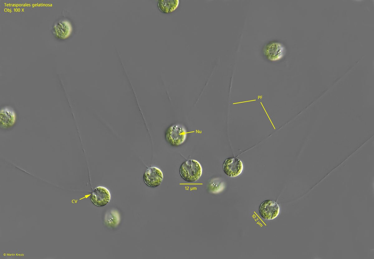
cells with 2 contractile vacuoles and 2 pseudoflagella
pseudoflagella extending beyond gelatinous mass
chloroplast cup-shaped, one pyrenoid
nucleus central
Tetraspora gelatinosa
I found Tetraspora gelatinosa for the first time in March 2019 in the Simmelried. I have not made any further findings of this tetrasporal alga so far.
The colony I found was about the size of a bunch of grapes and grew on a plant stem. It consisted of a very soft, gelatinous mass in which the cells were embedded both inside and also located on the surface. The spherical cells, with a diameter of 7–12 µm, have two conspicuous pseudoflagella, which can become very long and partly protrude from the surface of the colony (s. figs. 3 and 4). In addition, the cells have two contractile vacuoles apically, i.e., at the base of the pseudoflagella (s. fig. 4). The chloroplast is cup-shaped and sits posteriorly on the opposite side of the contractile vacuoles. The chloroplast also surrounds the centrally located nucleus (s. fig. 4). The cell wall is smooth and without visible structure.
Tetraspora gelatinosa occurs both in plankton and as a growth on aquatic plants. However, the alga requires very clear, cold, and clean water. With the onset of plankton formation in spring, it gradually disappears.
The similar species Tetraspora hexanematoidea does not have contractile vacuoles and has 2–6 pseudoflagella. This allows it to be reliably distinguished from Tetraspora gelatinosa.
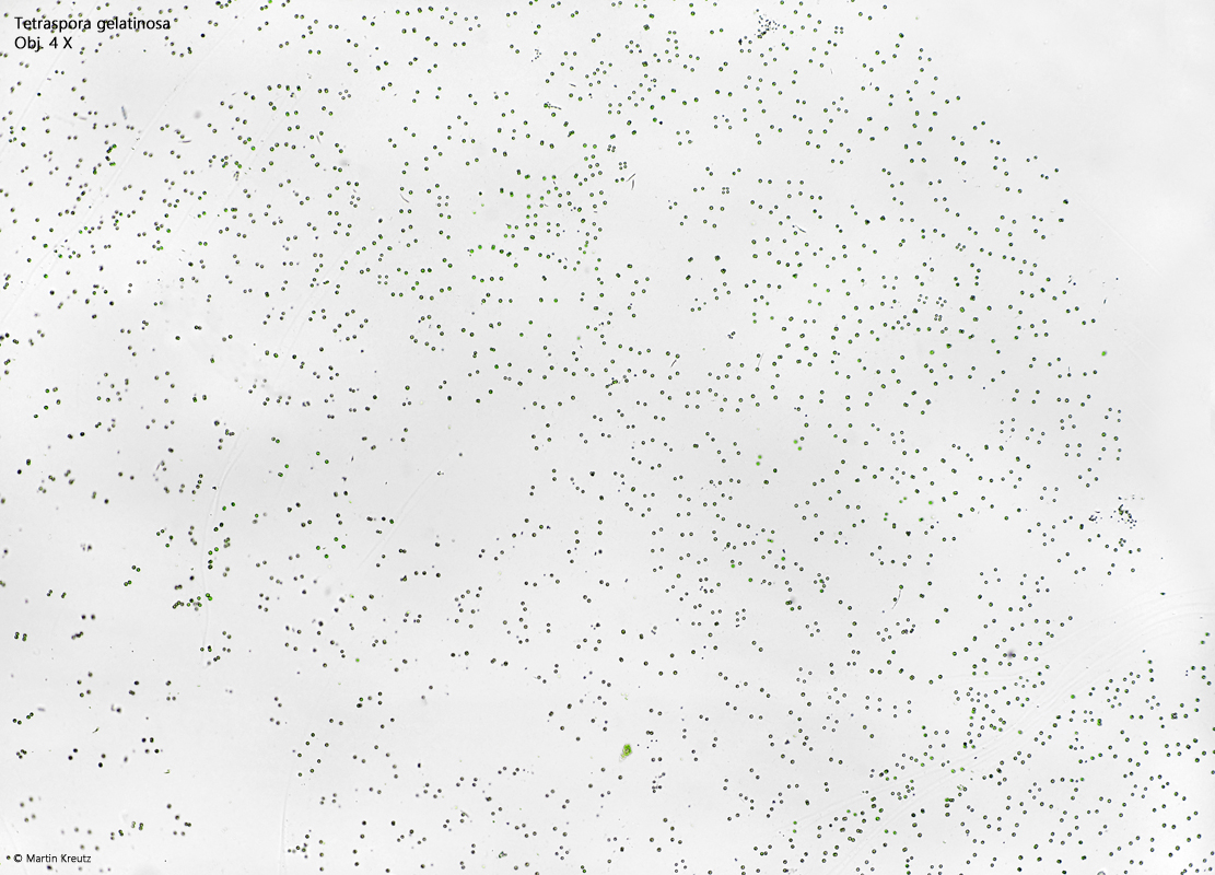
Fig. 1:Tetrapora gelatinosa. D = 3 cm (of colony). A small part of a squashed colony with several hundret cells. Obj. 4 X.
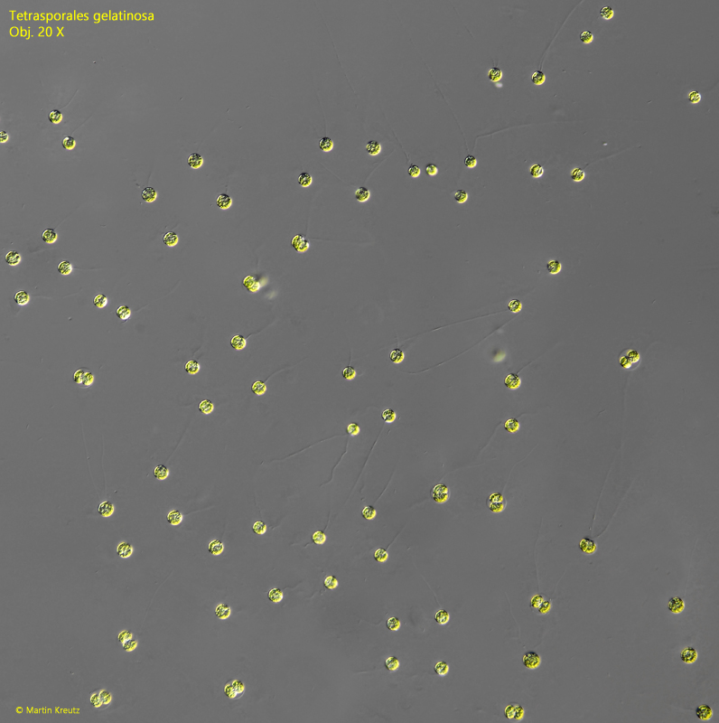
Fig. 2:Tetraspora gelatinosa. D = 3 cm (of colony). Focal plane on the surface of the squashed colony with the spherical cells empbedded in the gelatinous mass. Obj. 20 X.
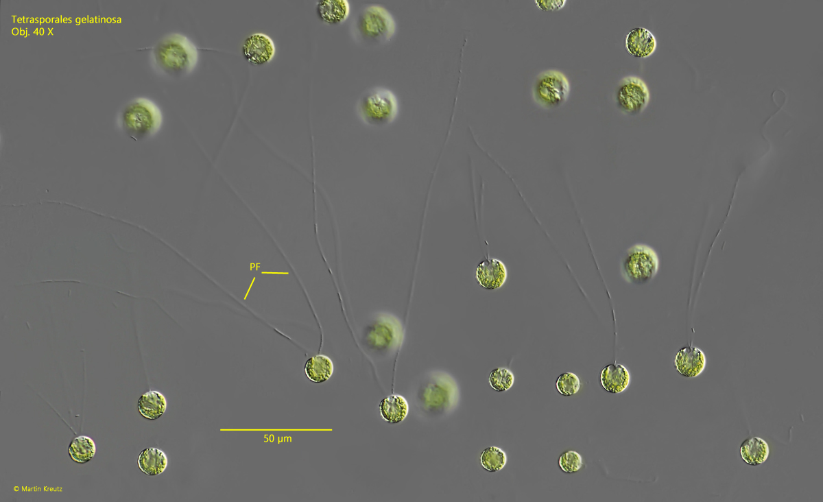
Fig. 3:Tetraspora gelatinosa. D = 9–12 µm (of cells). Some of the cells with each two, long pseudoflagella (PF). Obj. 40 X.
Fig. 4:Tetraspora gelatinosa. D = 9–12 µm (of cells). The spherical cells in detail. Each cell has two pseudoflagella (PF). Apically two contractile vacuoles (CV) are present, while the cup-shaped chloroplast is located basal. The nucleus (Nu) is located in the cavitiy of the chloroplast. Obj. 100 X.