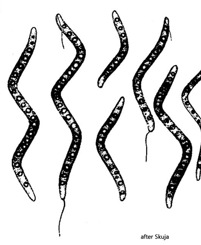
large sulphure globules in a row, scattered small globules
polar flagella at one or both cell ends
Thiospira dextrogyra
So far, I have only found Thiospira dextrogyra once, in December 2007, in the mud of Simmelried. The only accurate description of this sulfur bacterium seems to be that provided by Skuja (1956).
The species within the genus Thiospira are characterized by a spiral-shaped body and sulfur globules, which are always arranged in a row in the cells. In addition, the cells have flagella at one or both ends, which are often twisted into bundles.
The individual species within the genus Thiospira differ mainly in terms of cell diameter and length. The cells in my population had a diameter of 1.2–1.4 µm. This rules out the species Thiospira tenuis, which has a diameter of 0.8–1.0 µm, and Thiospira windradskyi, which has a diameter of 1.5–3.5 µm. The species Thiospira bipunctata has two conspicuously large sulfur globules and no globules arranged in a row. This leaves the species Thiospira dextrogyra, which is said to have a diameter of 1.0–1.3 µm. This corresponds to the diameter of the cells in my population. However, Skuja specifies a cell length of 15–35 µm, while the cells in my population were 28-53 µm long. Skuja also described the slightly longer variety Thiospira dextrogyra var. leptosoma, which is said to grow up to 40 µm long but has a diameter of only 0.7–0.9 µm. Although my specimens were slightly longer than those described by Skuja, the characteristics fit Thiospira dextrogyra better.
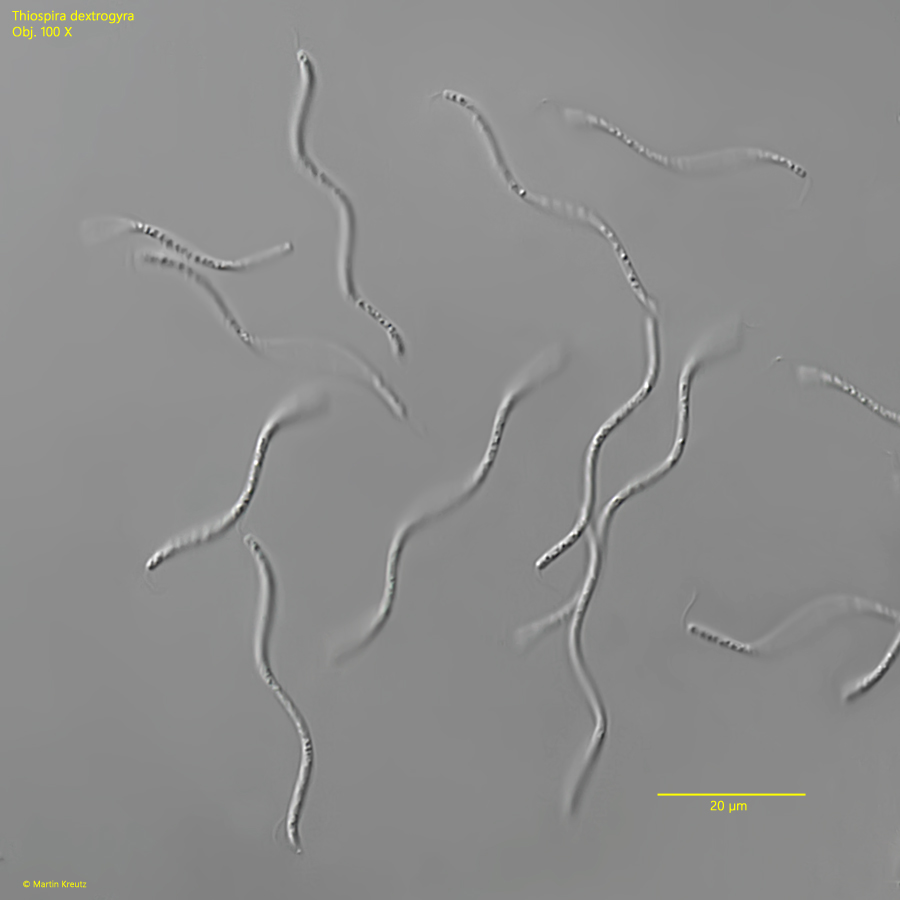
Fig. 1: Thiospira dextrogyra. L = 33–53 µm. Several freely swimming specimens. Obj. 100 X.
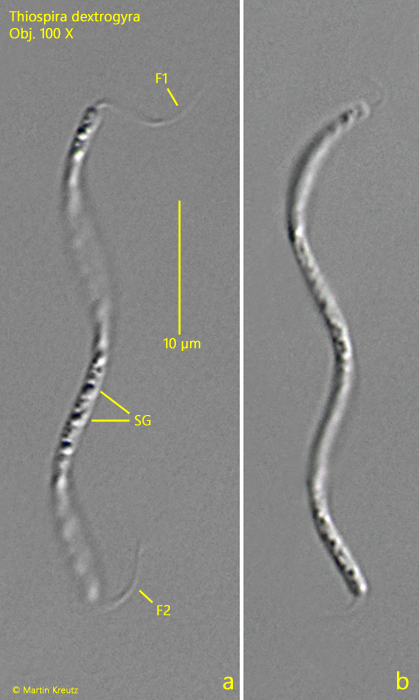
Fig. 1: Thiospira dextrogyra. L = 38–39 µm. Two specimens in detail. The specimens have a bundle of flagella at both ends (F1, F2). SG = sulphur globules. Obj. 100 X.