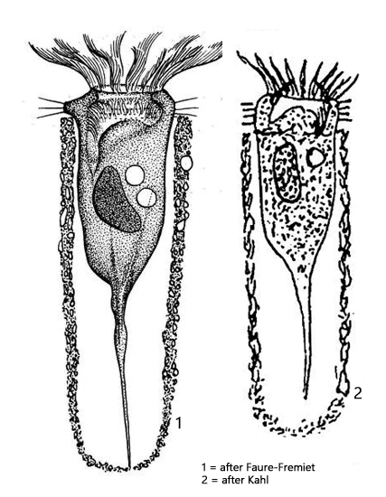
lorica soft, gelatinous, covered with detritus and diatoms
lorica up to a 280 µm long
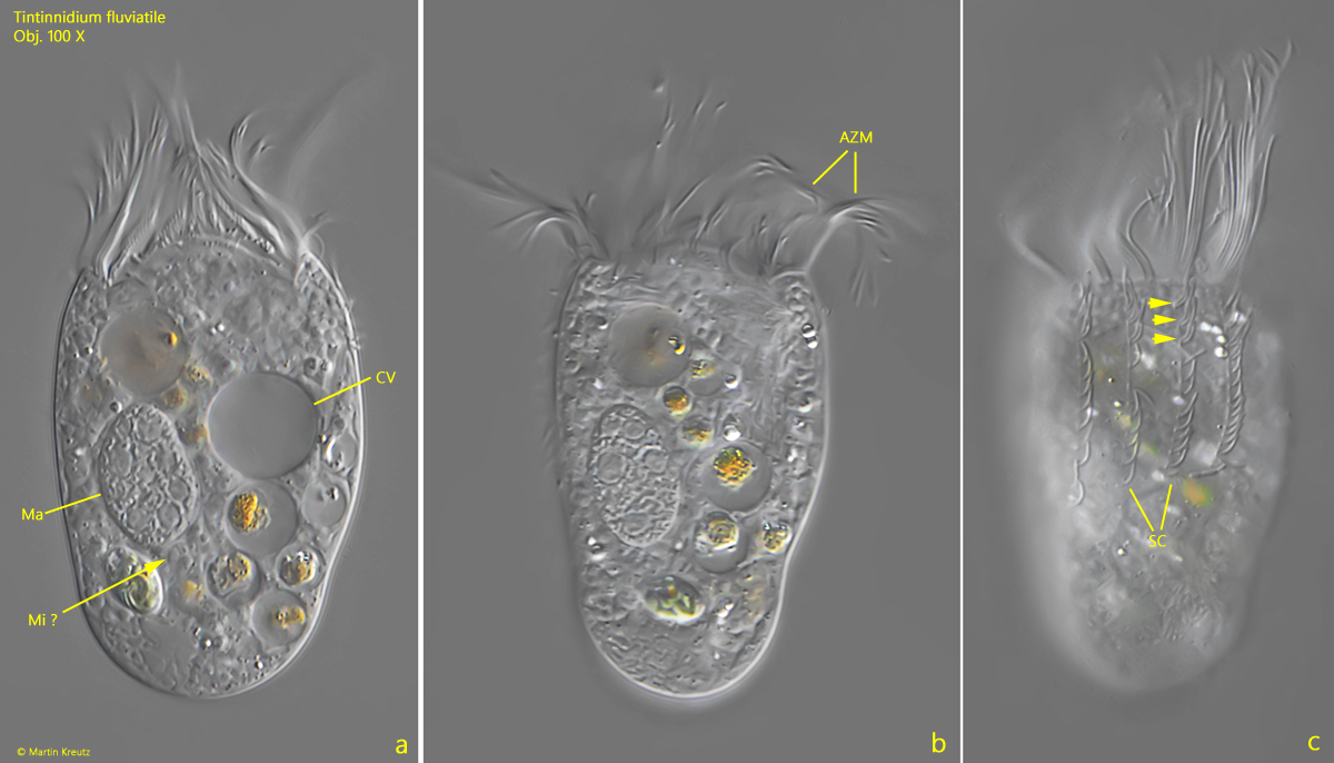
contractile vacuole near anterior end
macronucleus ellipsoid, near midbody
ellipsoidal micronucleus adjacent to macronucleus
oral apparatus antior with 12–14 adoral membranelles
usually 10 longitudinal ciliary row
three anterior cilia paired
planktonic lifestyle and sessile
Tintinnidium fluviatile
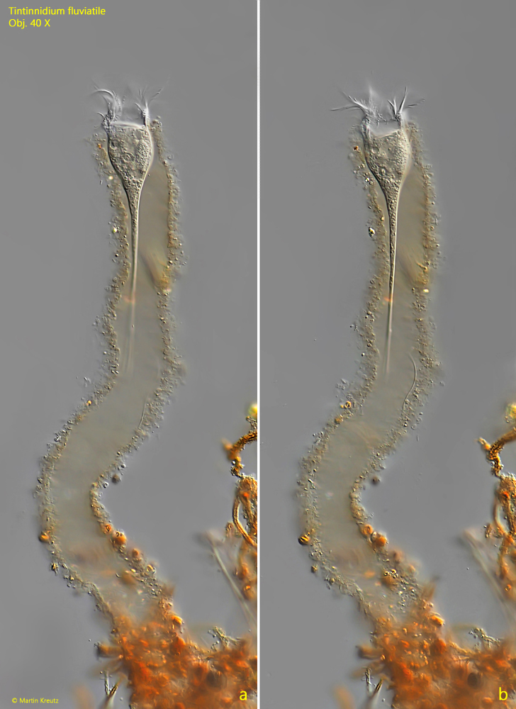
Tintinnidium fluviatile is a very common ciliate that I find in the plankton of all my sampling sites. However, sessile specimens are also found, whose loricae are considerably longer and covered with brownish, organic detritus. I usually find sessile specimens on the vessel walls of old samples or on the floating coverslip.
The loricae of Tintinnidium fluviatile can vary greatly in appearance, as they are covered with material from the ciliate’s habitat. This can be diatoms, sand, algae, or even detritus. The loricae can vary in length, as they are apparently constantly elongated during the ciliate’s lifetime.
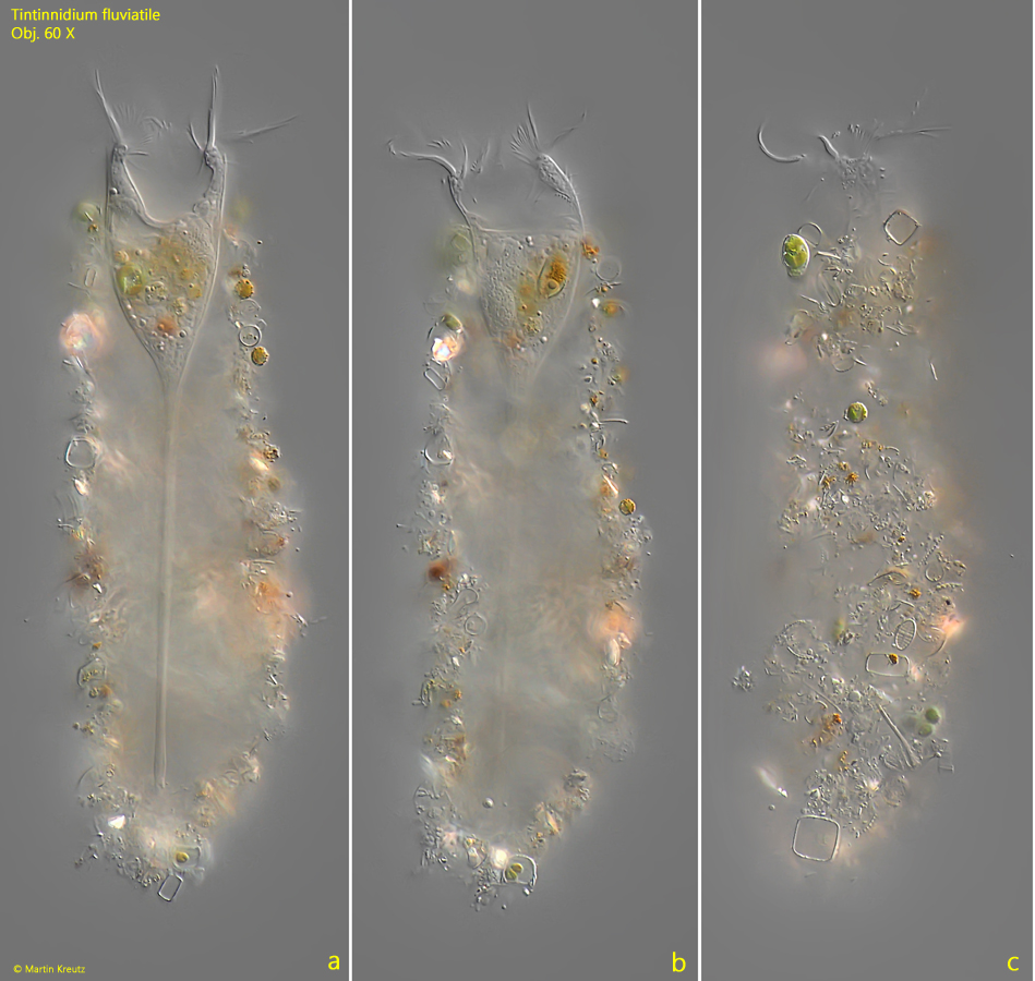
The specimens in my population were mostly 140–160 µm long, which allows for reliable differentiation from the similar species Tintinnidium pusillum, which has an average length of 50–80 µm. In addition, the loricae of Tintinnidium pusillum have a diameter significantly smaller than 30 µm.
Another important feature for the unambiguous identification of Tintinnidium fluviatile is the arrangement of the somatic cilia. These are 10 longitudinal rows of varying lengths that run across the body. Only the first three cilia at the anterior end of these rows are paired (s. fig. 4 c). The remaining cilia are single. This feature can be recognized by reducing the layer thickness. The specimens then leave the loricae and can be closer examined.
Fig. 1 a-b:Tintinnidium fluviatile. L = 166 µm. A sessile Specimen in a 355 µm long gelatinous lorica covered with orange-brownish detritus. The diameter of the lorica is 36 µm. Obj. 40 X.
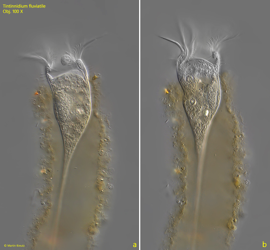
Fig. 2 a-b:Tintinnidium fluviatile. L = 166 µm. The same specimen as shown in fig. 1 a-b in detail. Obj. 100 X.
Fig. 3 a-c:Tintinnidium fluviatile. L = 146 µm. Three focal planes of a specimen with planktonic lifestyle. The lorica is 155 µm long and covered with diatoms, sand grains and small algae. Obj. 60 X.
Fig. 4 a-c:Tintinnidium fluviatile. L = 56 µm. A squashed specimen. Note the longitudinal rows of somatic cilia (SC). The first 3 cilia of these rows are paired (arrows). AZM = adoral zone of membranelles, CV = contractile vacuole, Ma = macronucleus, Mi ? = probably the micronucleus. Obj. 100 X.