I regularly find Tokophrya fasciculata as growth on the stems of the Yellow Water Lily (Nuphar lutea) in the pond of the waste disposal company Constance. On the stems this suctorian is difficult to study. Therefore, I cut some stems into pieces and place them with some water from the sampling site into a Petri dish. Then I place floating coverslips glasses on the surface. After 2–3 days I find specimens of Tokophrya fasciculata that have settled on the coverslip.
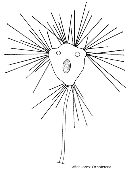
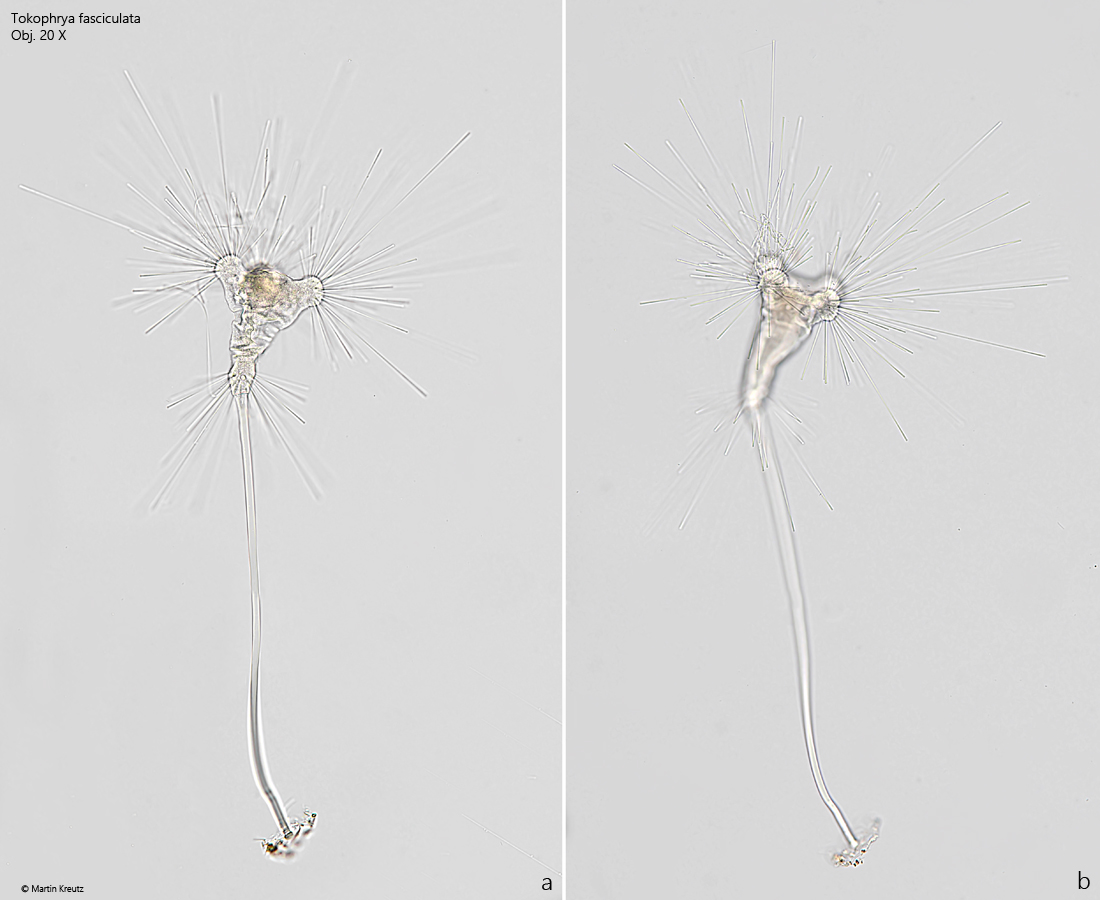
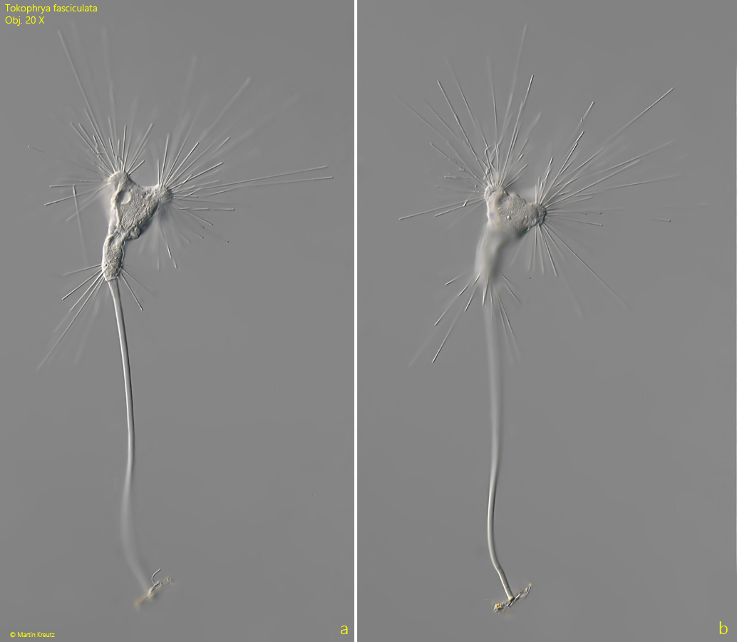
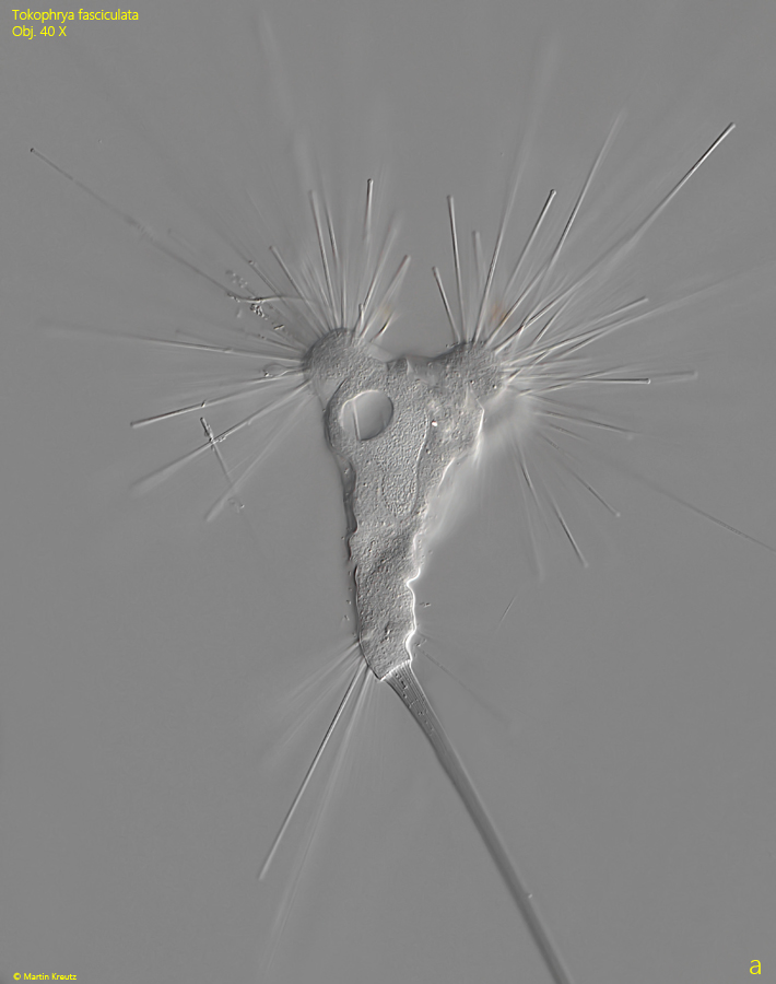
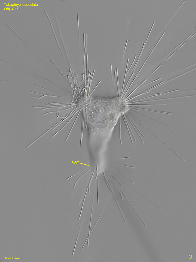
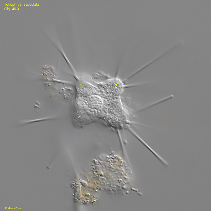
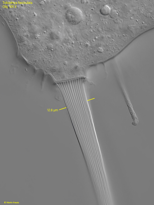
Tokophrya fasciculata is a fairly large suctorian and has striking features that make identification easy. Apically, the body has four bulges, from each of which a bundle of tentacles arises. In addition, Tokophrya fasciculata has, at the posterior end just before the base of the stalk, a wreath of tentacles that emerge from the body in a ring shape (s. figs 1 a-b and 3 b). The stalk formed by this suctorian is long and distinctly longitudinally striped (s. fig. 6).
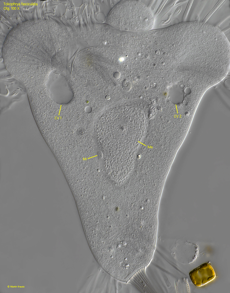
As further characteristics, Tokophrya fasciculata has two contractile vacuoles that lie near the apical bulges. The macronucleus lies centrally with an associated micronucleus (s. fig. 5).
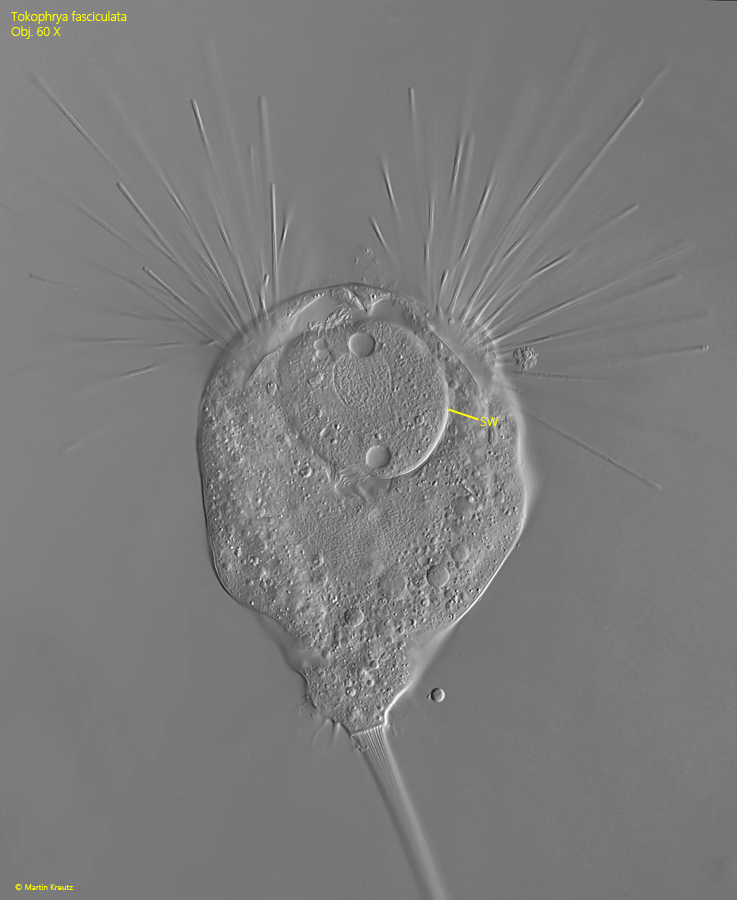
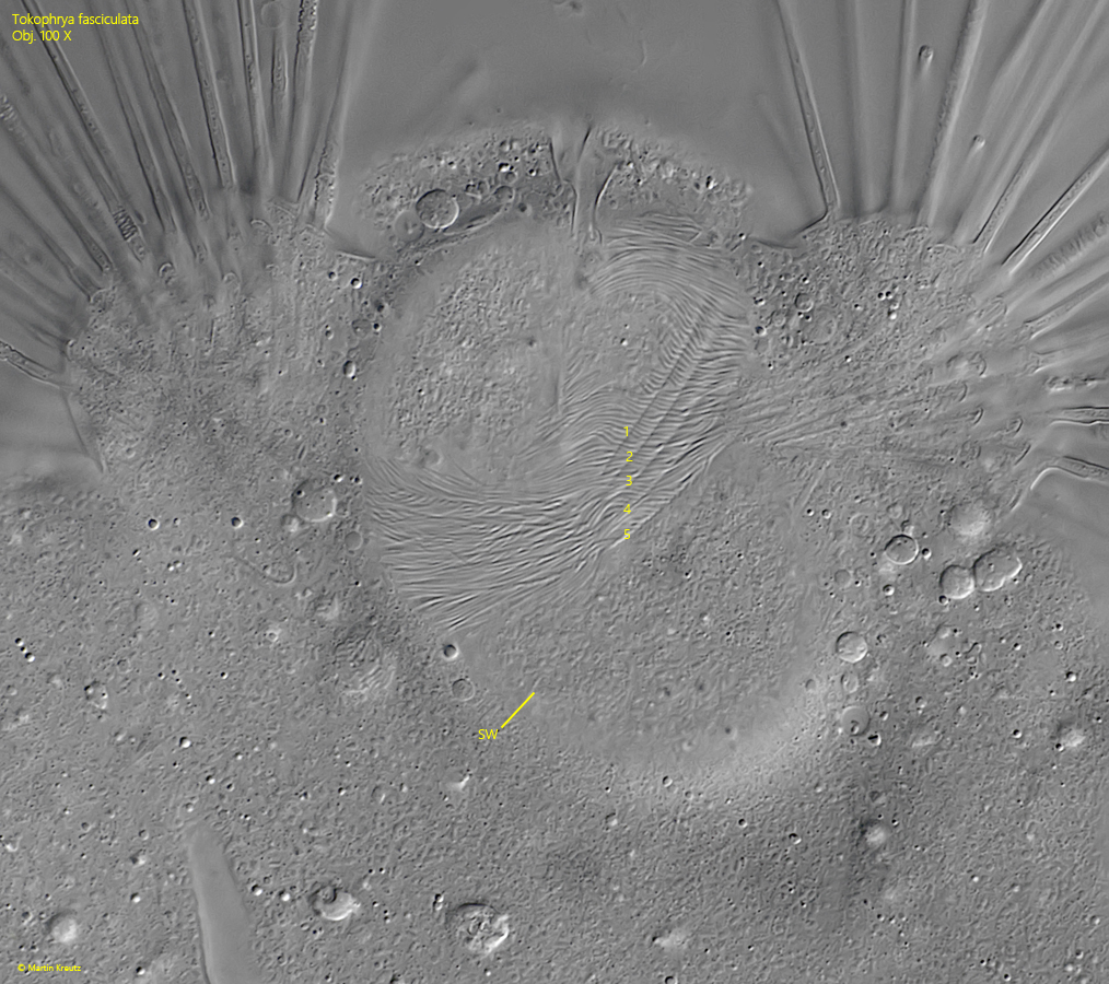
In some specimens I was able to observe the development of swarmers (s. fig. 10). They develop in an apical invagination and swim away after maturation. I was not able to find a freely swimming swarmer, but in the specimens developing in the depression I was able to observe 5 apical rows of cilia (s. fig. 11). In the literature available to me I have not found any description of the swarmer of Tokophrya fasciculata. Even the original describer Lopez-Ochoterena (1964) did not describe the swarmer.
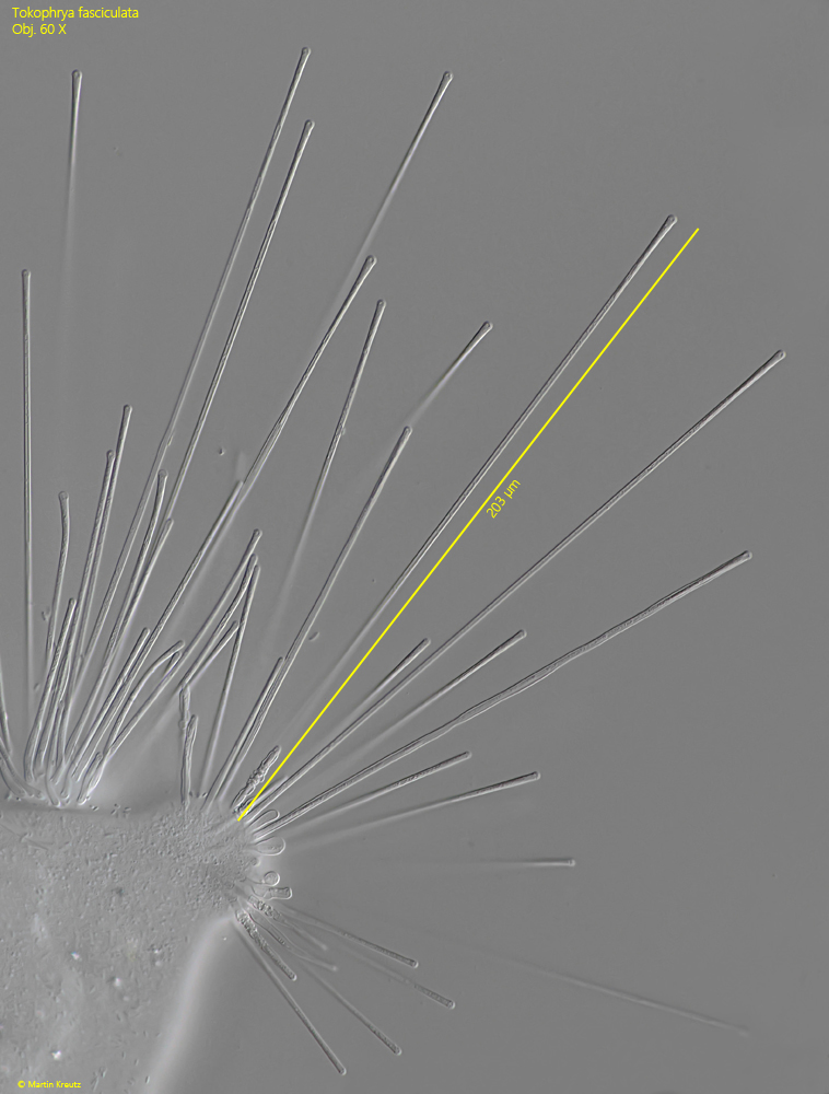
The specimens of my population had a body length of 130–160 µm, which is clearly longer than the 96–126 µm given by Lopez-Ochoterena (1964). The tentacles were also significantly longer, reaching up to 220 µm (s. fig. 7). I attribute this to the nutrient-rich habitat. Moreover, there are so far only a few records of Tokophrya fasciculata, which is why little is known about the variability of the species.
Fig. 1 a-b:Tokophrya fasciculata. L = 580 µm (with stalk, without tentacles). Two focal planes of an extended specimen in brightfield illuminaton. Apically four knobs with each one bundle of tentacles is present as well as a posterior wreath of tentacles. Obj. 20 X.
Fig. 2 a-b:Tokophrya fasciculata. L = 580 µm (with stalk, without tentacles). The same specimen as shown in fig. 1 a-b in DIC. Obj. 20 X.
Fig. 3 a-b:Tokophrya fasciculata. L = 152 µm (of body). Two focal planes of the body. Note the posterior wreath of tentacles (PWT). Obj. 40 X.
Fig. 4:Tokophrya fasciculata. Apical view on the four knobs (1–4) where the bundles of tentables arise. Obj. 40 X.
Fig. 5:Tokophrya fasciculata. A slightly squashed specimen. The two apical contractile vacuoles are visible (CV 1, CV 2) and the macronucleus (Ma) with an adjacent micronucleus (Mi) in the center. Obj. 100 X.
Fig. 6:Tokophrya fasciculata. The stalk with diameter of 12.8 µm has a distinct longitudinal striation. Obj. 100 X.
Fig. 7:Tokophrya fasciculata. One of the apical bundles of tentacles in detail. They have a length of about 200 µm. Obj. 40 X.
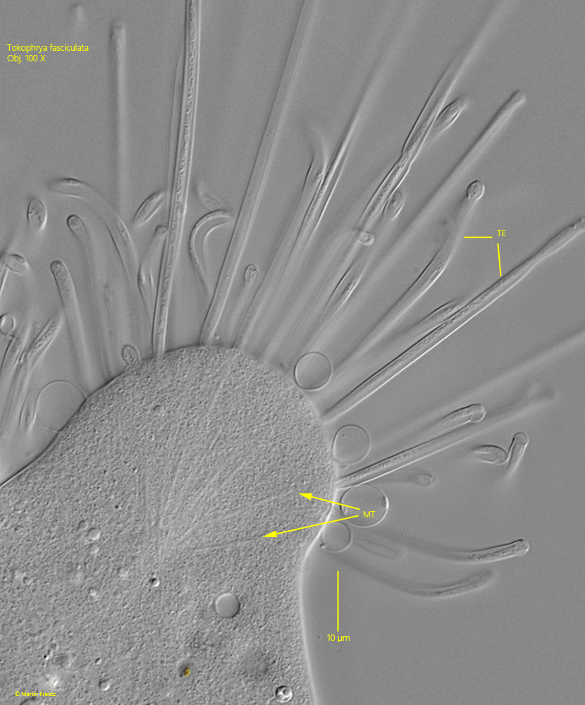
Fig. 8:Tokophrya fasciculata. In the cytoplasm of the knobs bundles of microtubules (MT) are visible where the apical tentacles (TE) arise. They continue into the tentacles. Obj. 40 X.
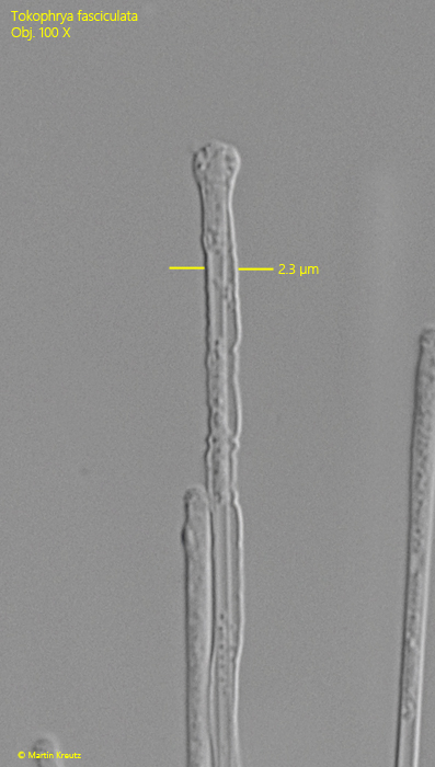
Fig. 9:Tokophrya fasciculata. One of the tentacles in detail. They have a diameter of 2.2–2.5 µm. Obj. 100 X.
Fig. 10:Tokophrya fasciculata. L = 130 µm (of body). A specimen with an spherical swarmer (SW) in an apical invagination. The two contractile vacuoles as well as the macronucleus of the swarmer are visible. Obj. 100 X.
Fig. 11:Tokophrya fasciculata. Focal plane on the 5 apical rows of cilia (1-5) of the swarmer (SW). Obj. 100 X.