body shape elongate, flask-like, dorso-ventrally flattened
bent anterior neck region
length 250–480 µm, width 40–90 µm
oral bulge with 2 types of extrusomes
type 1 extrusomes filiform, 35–65 µm long
type 2 extrusomes rod-shaped, 5–8 µm long
cell covered by mucuos layer (6–7 µm) with lepidosomes
4 types of lepidosomes, 1.6–5 µm
two macronuclear nodules connected vias a strand (hard to see)
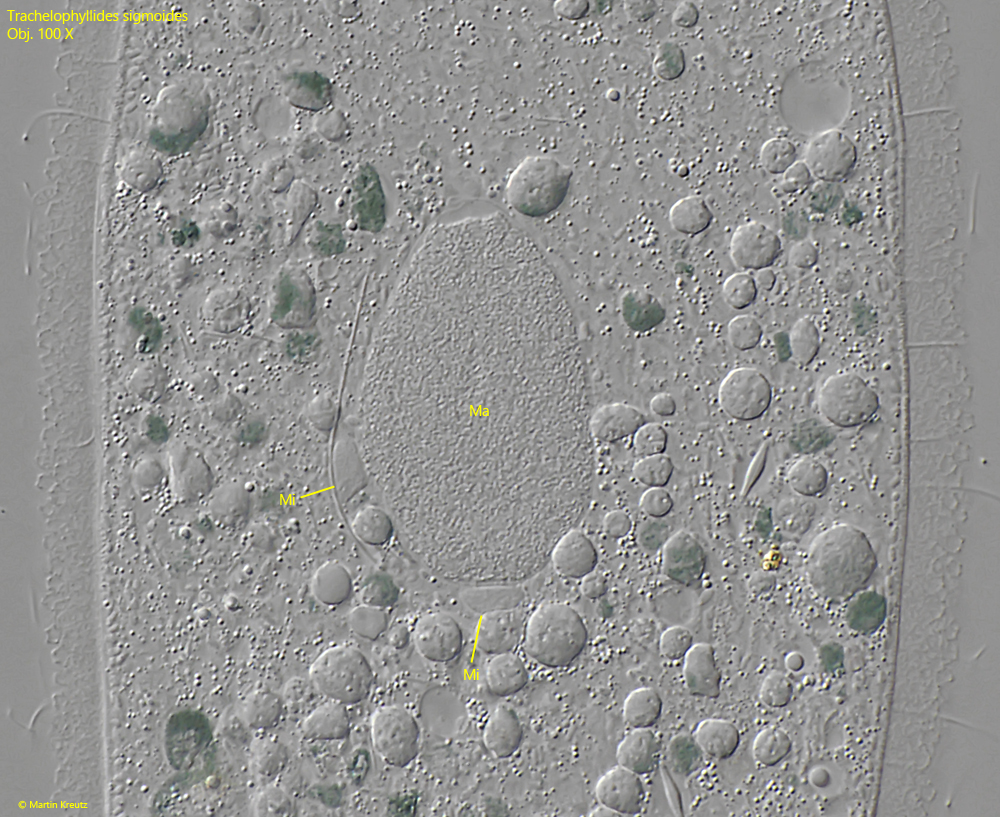
each macronuclear nodule with several micronuclei (5–6 µm)
cytoplasm sometimes green due to symbiotic algae
contractile vacuole terminal
Trachelophyllides sigmoides
I find Trachelophyllides sigmoides rarely, but regularly in the Simmelried. I have not yet been able to find this ciliate in my other sampling sites. The specimens are usually found in samples of floating and decomposing plant masses. In old samples (2–3 weeks) they can be found at the bottom of the container.
Trachelophyllides sigmoides was originally described as Trachelophyllum sigmoides by Kahl in 1926. In 2016 Foissner then carried out very thorough studies (electron microscopically) on the lepidosomes of the trachelophyllid ciliates, which are found in the mucuos sheath of these ciliates. He was able to determine significant differences in the shape of the lepidosomes and subsequently introduced new genera. As a result of his investigations, he transferred Trachelophyllum sigmoides to the new genus Trachelophyllides.
Trachelophyllides sigmoides is a very large ciliate (usually 300-400 µm) and is very transparent and easy to observe due to its strong dorso-ventral flattening (s. fig. 1 a-c). In addition, Trachelophyllides sigmoides moves only slowly. It glides on the substrate and rarely swims freely. In the specimens it is immediately recognizable by its size and the always bent or curved neck, which ends in the oral bulge. The oral bulge is equipped with two types of extrusomes that can be easily recognized. The short extrusomes of type II form a short, sharply defined fringe at the distal end of the oral bulge. The long (up to 65 µm) extrusomes of type 1 lie as a dense bundle underneath (s. fig. 2).
Even at low magnifications it is clearly visible that Trachelophyllides sigmoides is covered by a distinct mucuos sheath. This layer has an extremely complex structure because lepidosomes are embedded in it. At low magnification, the mucuos sheath therefore appears rough. At high magnification, however, the lepidosomes can be recognized (s. figs. 3 and 4). In the case of Trachelophyllides sigmoides, there are 4 different lepidosomes, which can only be distinguished under an electron microscope. They are bowl-shaped or mushroom-shaped and have window-like openings, giving them a polygonal surface (s. drawing above). These “windows” can also be seen under a light microscope at maximum magnification (s. fig. 4). The purpose of the mucuos sheath the complex structure of the lepidosomes is still unclear.
Sometimes green specimens of Trachelophyllides sigmoides are also found because they contain Chorella-type algae (s. fig. 6). They are not found in food vacuoles and are permanently present. However, if the ciliate is burst and the algae are released, they quickly bleach out. Foissner (2016) has therefore suggested that these are not symbiotic algae but cleptoplasts, i.e. algae stolen from prey ciliates that are kept alive.
Fig. 1 a-c:Trachelophyllides sigmoides. L = 390 µm. A freely swimming specimen from right. Note the mucuos sheath (MS) with lepidosomes covering the cell. CV = contractile vacuole, Ma = macronuclear nodule, Mi = micronuclei, OB = oral bulge. Obj. 40 X.
Fig. 2:Trachelophyllides sigmoides. The oral bulge in detail with the two types of extrusomes (EX I, EX II). The mucuos sheath of this specimen is 7.7 µm thick. Obj. 40 X.
Fig. 3:Trachelophyllides sigmoides. The lepidosomes (LE) in the mucuos sheath of a squashed specimen in lateral view. They have a polygonal structure. Obj. 100 X.
Fig. 4:Trachelophyllides sigmoides. The lepidosomes (LE) in the mucuos sheath of a squashed specimen in frontal view. The most lepidosomes appear pentagonal or hexagonal in optical section. In one case (arrow) the “windows” of the polygonal structure of the lepidosomes are visible. Obj. 100 X.
Fig. 5:Trachelophyllides sigmoides. One of the two macronuclear nodules (Ma) with two adjacent micronuclei (Mi). Obj. 60 X.
Fig. 6:Trachelophyllides sigmoides. L = 250–266 µm. Two specimens start to create a bridge of cytoplasma between their oral bulges to start the conjugation. Obj. 40 X.