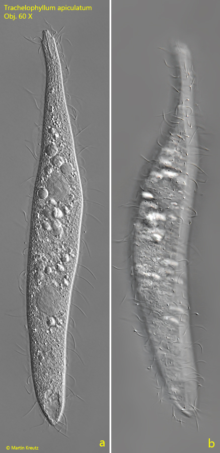
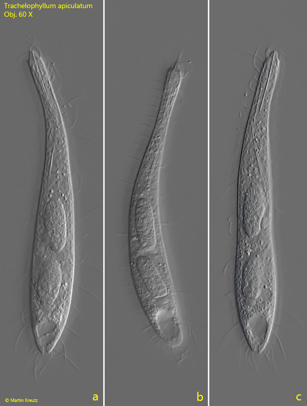
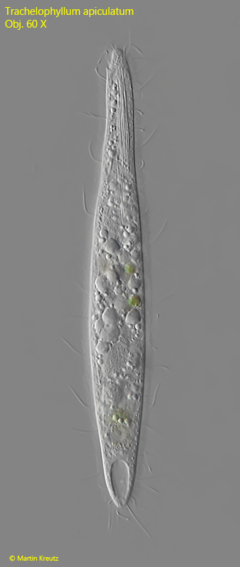
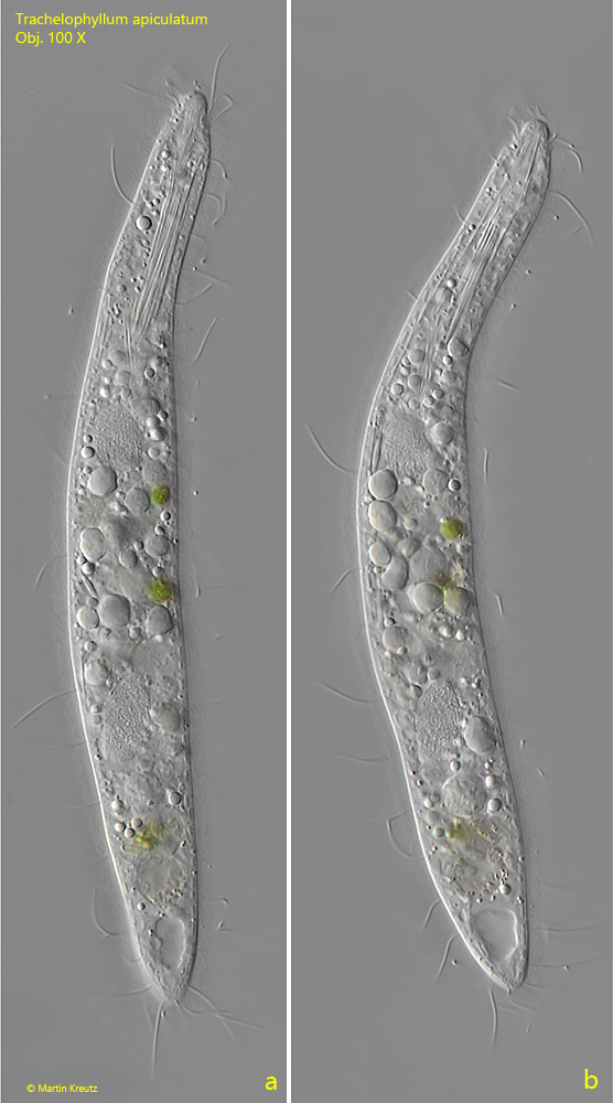
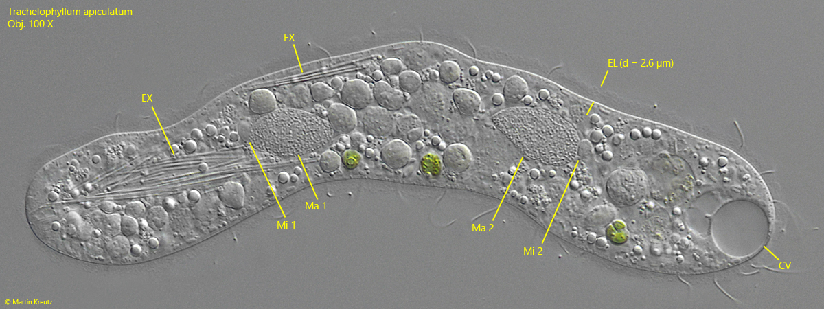
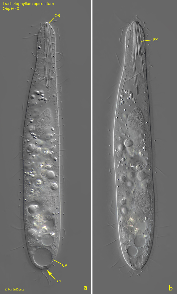
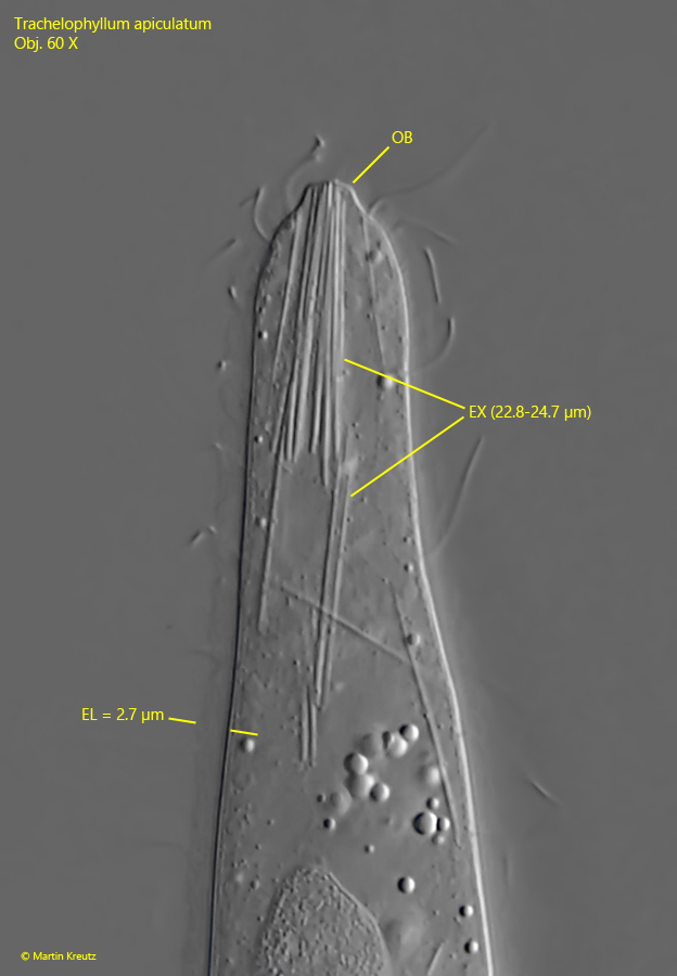
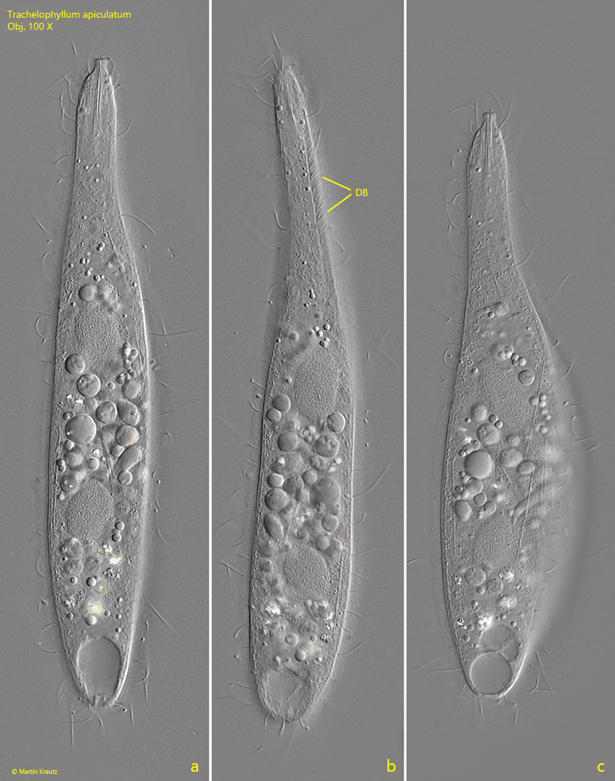
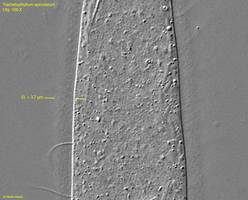
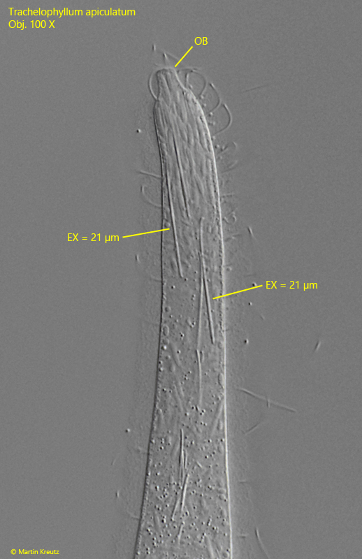
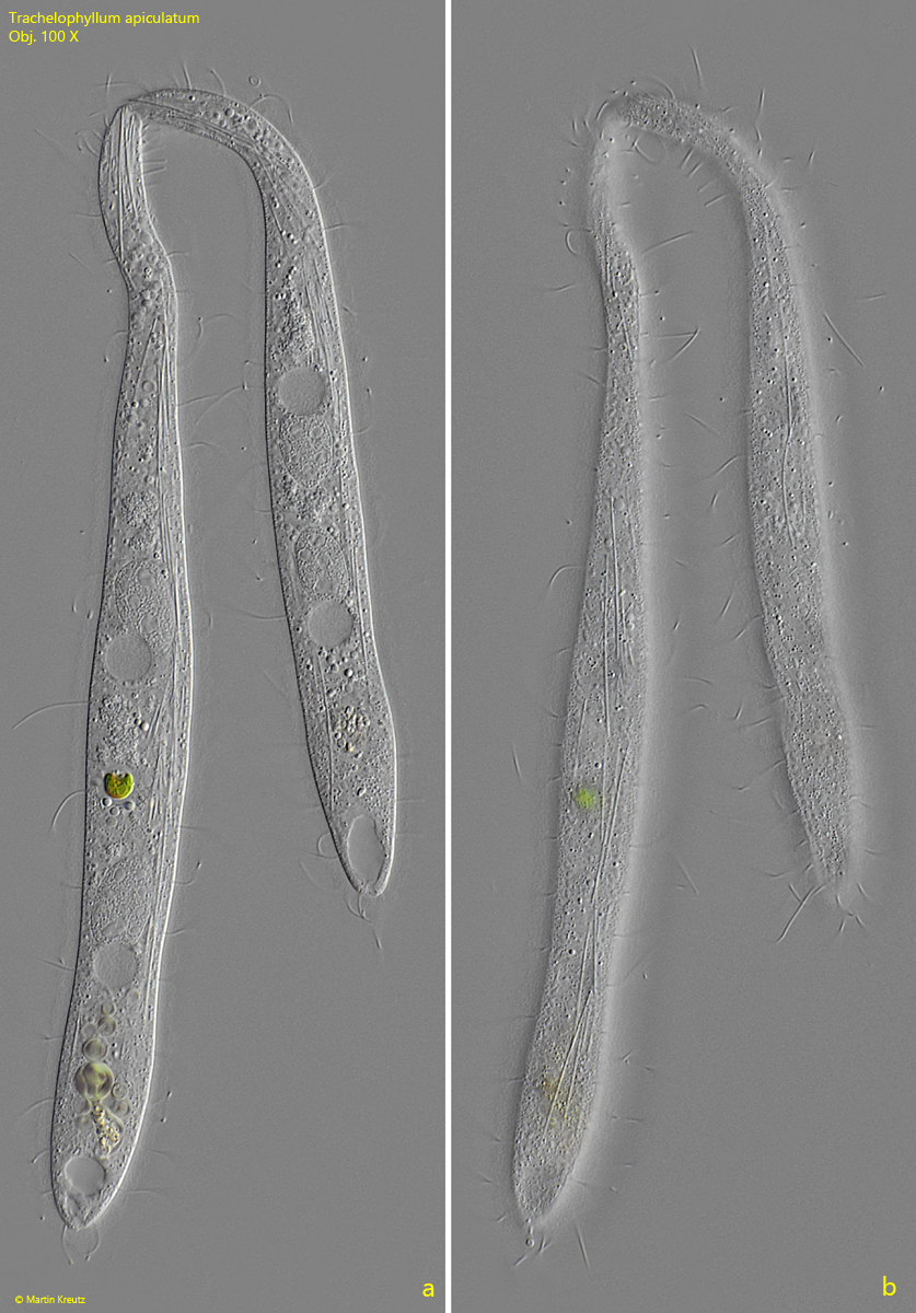
Trachelophyllum apiculatum
(Perty, 1852) Claparède & Lachmann, 1859
Most likely ID: n.a.
Â
Synonym: n.a.
Â
Sampling location: Simmelried
Â
Phylogenetic tree: Trachelophyllum apiculatum
Â
Diagnosis:Â
- body slenderly club-shaped or fusiform, flattened
- oral bulge conical or cylindrical
- length 100–200 µm, width 15–30 µm
- two widely separated, ellipsoid macronuclei
- each macronucleus with one micronucleus
- 10–25 longitudinal rows of cilia
- extrusomes rod-shaped, 10–20 µm long, bundled in cytopharynx
- cells covered by epicortical layer with lepidosomes
- dorsal brush three-rowed
- contractile vacuole terminal
- excretion pore terminal
So far, I have found Trachelophyllum apiculatum exclusively in the Simmelried. Mostly in old samples with few plant remains.
Â
The literature on Trachelophyllum apiculatum is quite heterogeneous, and there are different reports regarding the size, the extrusomes, or the existence of the epicortical layer. Kahl (1935) provides only a very brief description. He gives a length of 125–150 µm and does not mention the extrusomes or an epicortical layer. Foissner published a redescription in 1983. Here, Foissner gives a body length of only 90–110 µm and 13 µm length for the rod-shaped extrusomes. His drawing from 1983 (s. drawing a, above) shows a very slender specimen without an epicortical layer. In 1995, Foissner, Berger, Blatterer, and Kohmann published an further description of Trachelophyllum apiculatum, in which the authors described the species without an epicortical layer, with a length of 90–180 µm and 15 µm long extrusomes. Seven years later, in 2002, Foissner, Berger, and Agatha published the results of their investigations on a population of Trachelophyllum apiculatum from Venezuela. In this publication, the authors were able to detect the epicortical layer, into which intricately shaped scales (lepidosomes) are embedded. The size of the specimens is given as 100-200 µm, and the length of the extrusomes as 10-20 µm. The epicortical layer is described as very hyaline with a thickness of 1–2.5 µm.
Â
This rough overview of the available descriptions of Trachelophyllum apiculatum gives an impression, how the data has changed over time with the number of specimens and populations examined. Foissner, Berger, and Agatha (2002) therefore assume that the species is very variable and that there may possibly be a species complex behind it.
Fig. 1 a-b: Trachelophyllum apiculatum. L = 218 µm. A large specimen found in November 2014 in the Simmelried. Obj. 60 X.
Fig. 2 a-c: Trachelophyllum apiculatum. L =107 µm. A small specimen with large macronuclei found in April 2021 in the Simmelried. Obj. 60 X.
Fig. 3: Trachelophyllum apiculatum. L =150 µm. A freely swimming, spindle-shaped specimen. Obj. 60 X.
Fig. 4 a-b: Trachelophyllum apiculatum. L =150 µm. The same specimen as shown in fig. 3 at higher magnification. Obj. 100 X.
Fig. 5: Trachelophyllum apiculatum. The spuashed specimen as shown in fig. 4 a-b. Note the delicate epicortical layer with a granular appearance. The two macronuclei (Ma 1, Ma 2) have each one micronucleus (Mi 1, Mi 2). EX = rod-shaped extrusomes. Obj. 100 X.
Fig. 6 a-b: Trachelophyllum apiculatum. L =160 µm. A transparent specimen with a budle of extrusomes (EX), parallel arranged in the cytopharynx. The distal end of the extrusomes reach to the top of the oral bulge (OB). At the posterior end the excretion pore (EP) of the contractile vacuole (CV) is visible. Obj. 60 X.
Fig. 7: Trachelophyllum apiculatum. A crop of fig. 1 b. The rod-shaped extrusomes (EX) have length of 22.6–24.7 µm) and the epicortical layer (EL) is 2.7 µm thick. Obj. 60 X.
Fig. 8 a-c: Trachelophyllum apiculatum. L =145 µm. A slightly squashed specimen found in October 2021 in the Simmelried. Note the paired cilia of the dorsal brush (DB). Obj. 100 X.
Fig. 9: Trachelophyllum apiculatum. The epicortical layer (EL) with the thickness of 3.7 µm in a squashed specimen. The granular appearance is caused by embedded lepidosomes with a complex structure. The shape of the lepidosomes cannot be resolved in the light microscope. Obj. 100 X.
Fig. 10: Trachelophyllum apiculatum. The conical oral bulge (OB) of a slightly squashed specimen in detail. The extrusomes (EX) of this specimen have a length of 21 µm. Obj. 100 X.
Fig. 11 a-b: L = 173 + 134 µm. Trachelophyllum apiculatum. Two specimens in conjugation. Obj. 100 X.