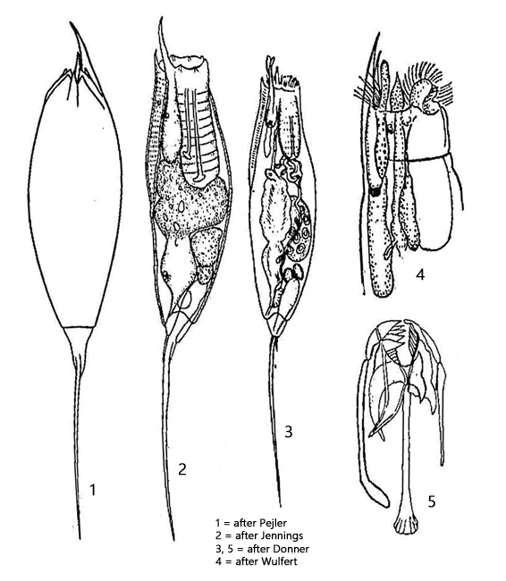
dorsal two inconspicuous keels and ribbon of stripes
length 300–575 µm (with toes)
one dorsal eyespot
stomach and intestine with fat spherules
retrocerebral organ with elongated sac
left toe, almost body length
right toe, short and curved
5–7 very short, secondary toes (hard to see)
cuticle often colored yellowish or reddish
Trichocerca longiseta
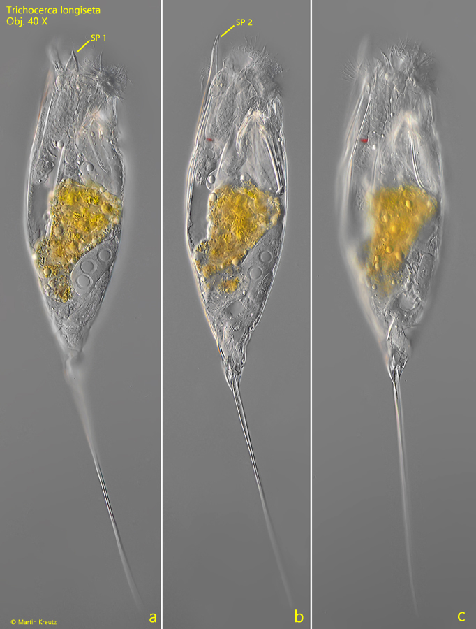
So far, I have found Trichocerca longiseta exclusively in the Simmelried among floating plants. With an average length of 400–500 µm and the conspicuous long left toe, the specimens in the samples are very noticeable. Most specimens in my population were colorless, yellowish, and sometimes intensely orange (s. fig. 3).
Besides the long left toe, two spines on the apical margin are an important identification feature. The more slender spine is about twice as long as the smaller and broader spine (s. figs. 1a and 1 b). Often, the stomach of the specimens is conspicuously yellow, orange, green, or sometimes also red. There is no information in the literature about the diet of Trichocerca longiseta, but it is suspected that it feeds on dinoflagellates, chrysophytes, and flagellated algae, which also inhabit the water plants.
Fig. 1 a-c:Trichocerca longiseta. L = 412 µm (with toes). Different focal planes of a specimen from right. Obj. 40 X.
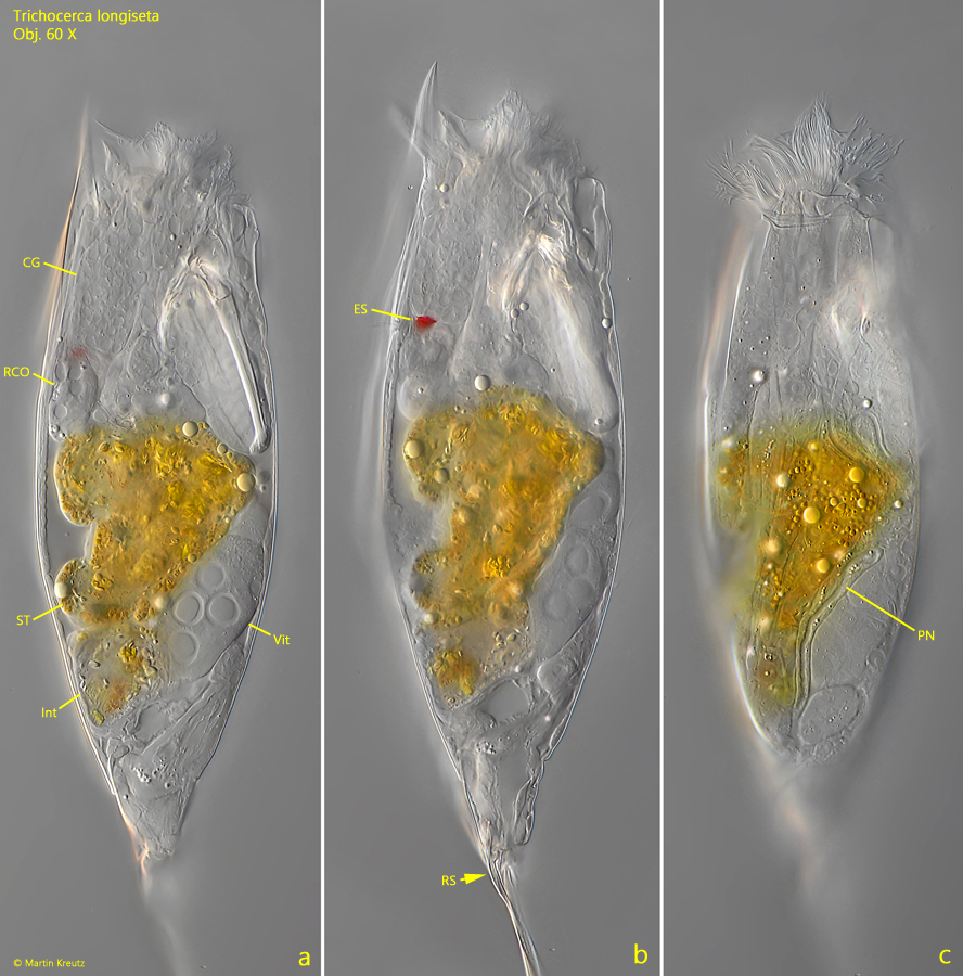
Fig. 2 a-c:Trichocerca longiseta. L = 412 µm (with toes). The body of the same specimen as shown in fig. 1 a-c in detail. CG = cerebral ganglion, ES = eyespot, Int = intestine, PN = protonephridium, RCO = retrocerebral organ, RS = short right toe, ST = stomach, Vit = vitellarium. Obj. 60 X.
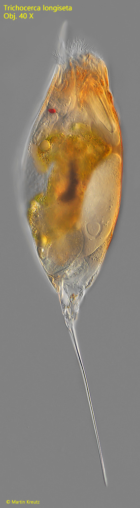
Fig. 3:Trichocerca longiseta. L = 472 µm (with toes). A second, intensely orange colored specimen. Obj. 40 X.
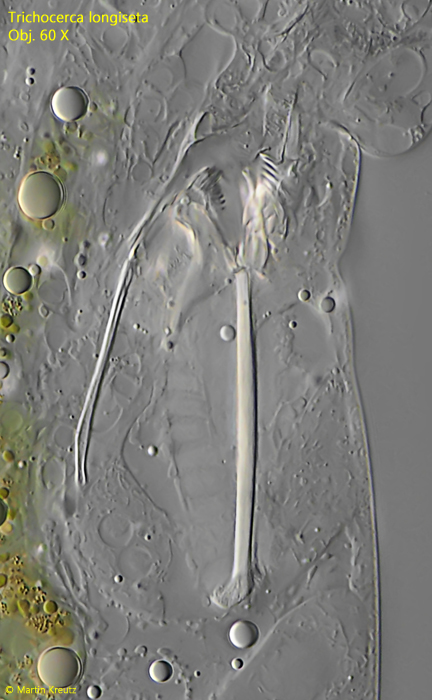
Fig. 4:Trichocerca longiseta. The trophy in a slightly squashed specimen. Obj. 60 X.
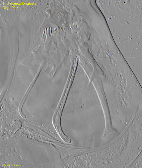
Fig. 5:Trichocerca longiseta. The trophi in a strongly squashed specimen. Obj. 100 X.