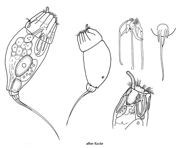
left toe one half of body length, ventrally curved
three secondary toes
toes bend ventrally in contracted specimens
lateral antennae short, near foot
eyespot in at dorsal end of cerebral ganglion
Trichocerca pusilla
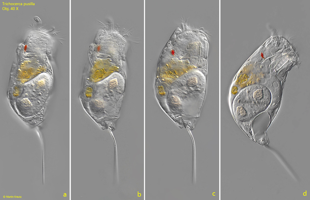
So far, I have found Trichocerca pusilla exclusively in the pond of the waste disposal company Constance. All specimens shown below come from plankton samples taken between May and September 2020.
In the fresh samples, the specimens of Trichocerca pusilla stand out due to their delicate shape. This is also due to the long, left main toe, which takes up about half of the body length. It is thin and evenly curved backward. Only in squashed specimens can one see that three additional secondary toes are present (s. fig. 7).
A key characteristic of Trichocerca pusilla is the position of the eyespot on the cerebral ganglion. It is located almost at the end of the cerebral ganglion on the dorsal side (s. fig. 6). Additionally, the apical margin of the cuticle shows a characteristic longitudinal folding when the specimens contract (s. fig. 4). In extended specimens, a curved palp organ as part of the corona can be clearly seen (s. fig. 2 b).
The similar species Trichocerca stylata is stouter with shorter toes, which make up only a quarter or a fifth of the body length. Furthermore, the eyespot in Trichocerca stylata is located in the middle of the cerebral ganglion and not at the end.
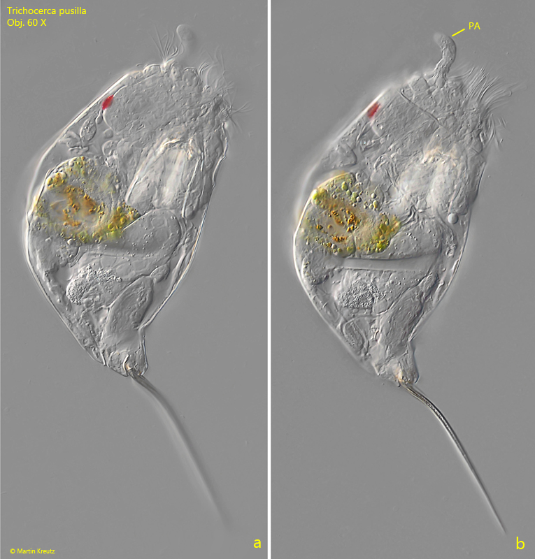
Fig. 2 a-b:Trichocerca pusilla. L = 172 µm (with toes). A slightly squashed second specimen from right. Note the ventrally bent over toes and the curved palp organ (PA). Obj. 60 X.
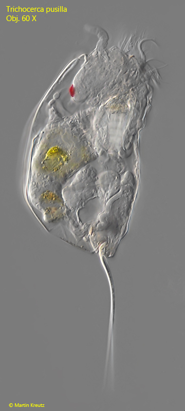
Fig. 3:Trichocerca pusilla. L = 160 µm (with toes). A third specimen from right. Obj. 60 X.
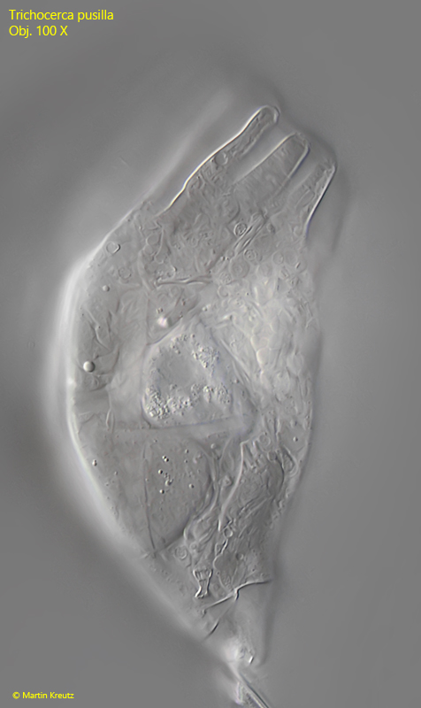
Fig. 4:Trichocerca pusilla. The folded apical margin of a contracted specimen. Obj. 100 X.
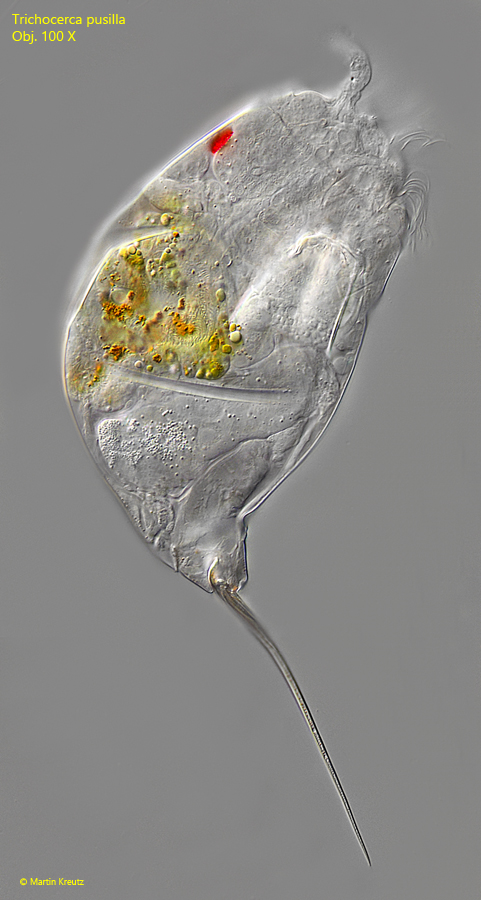
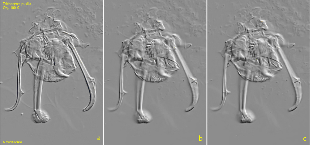
Fig. 5:Trichocerca pusilla. L = 175 µm (with toes). Total view of the specimen as shown in fig. 2 a-b at hight magnification. Obj. 100 X.
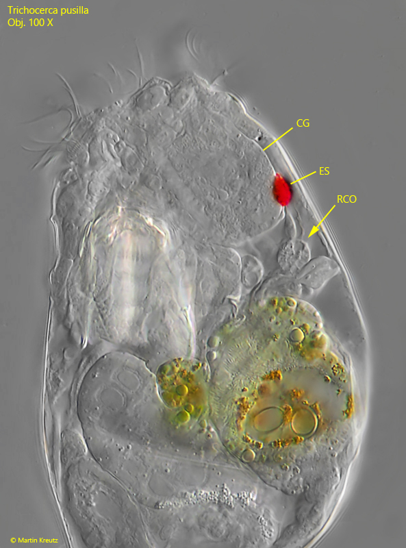
Fig. 6:Trichocerca pusilla. The head region in detail. The eyespot (ES) is attached to the end of the cerebral ganglion on the dorsal side. Below the almost spherical retrocerebral organ( RCO) is visible. Obj. 100 X.
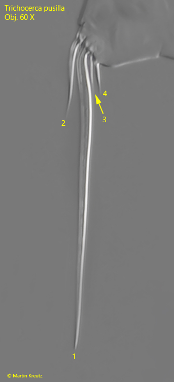
Fig. 7:Trichocerca pusilla. The main toe (1) and the three secondary toes (2-3) in a squashed specimen. Obj. 60 X.
Fig. 8 a-c:Trichocerca pusilla. Three focal planes of the trophi in a strongly squashed specimen. Obj. 100 X.
Fig. 9 a-c:Trichocerca pusilla. The trophi of a second specimen. Obj. 100 X.