retrocerebral organ divided in gland and reservoir
foot two-jointed
toes short of unequal length
Trichocerca similis
So far I have only found Trichocerca similis in the plankton of two sampling sites. Both waters are stocked with fish and are comparatively highly eutrophic. In general, however, I only rarely find Trichocerca similis.
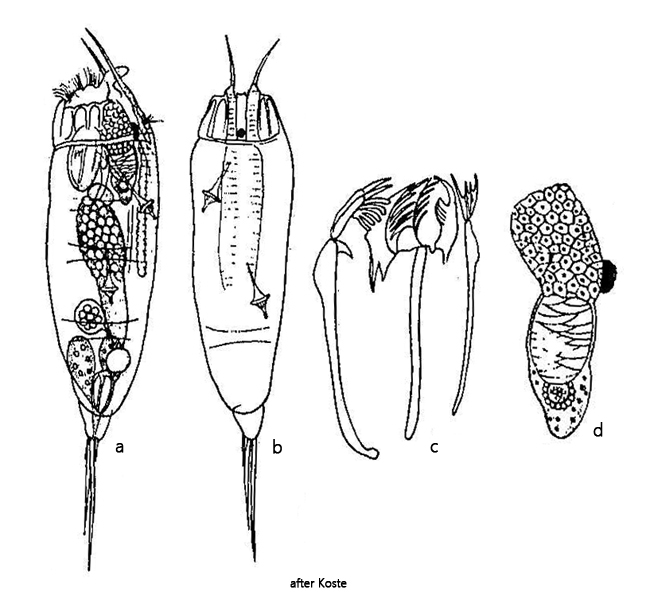
Trichocerca similis can be easily recognized by the two apical spines of equal length (s. fig. 2 a) and the rather short toes. At higher magnification, a striped band can also be seen on the dorsal side (s. fig. 2 b).
Another characteristic feature is the large retrocerebral organ, which is located below the eyespot (s. fig. 3). It is so conspicuous that it was reproduced by Koste (1978) in a separate drawing (s. drawing d above). It is clearly divided into a distal gland and an overlying reservoir filled with a layered mass (s. fig. 4). The function of this organ can be easily understood by this clear division.
Several gland cells are arranged around a collecting vacuole. They release a gelatinous glandular secretion into this via small vesicles. It is easy to see how the vesicles are arranged around the collecting vacuole (s. fig. 5). When the collecting vacuole is filled, the contents empty into the reservoir above. The secretion does not mix with the secretion already contained there, resulting in a layered structure that can be clearly seen (s. fig. 5).
The composition and function of the secretion formed is unknown. It is passed through a channel from the reservoir to the apex and excreted there. The secretion may contain enzymes that are mixed with the food that is swirled in to aid digestion.
Fig. 1 a-b:Trichocerca similis. L = 226 µm. A freely swimming specimen from right. Obj. 60 X.
Fig. 2 a-c:Trichocerca similis. L = 226 µm. The slightly squashed specimen as shown in fig. 1 a-b from right. Note the two apical spines (AS) of equal length and the dorsal band of stripes (BS). Obj. 40 X.
Fig. 3:Trichocerca similis. L = 240 µm. A second, slightly squashed specimen from right. Note the large retrocerebral organ (RCO) below the eyespot (ES). AS = apical spines, BL = bladder, GG = gastric gland, ST = stomach. Obj. 60 X.
Fig. 4:Trichocerca similis. The retrocerebal organ is divided in a gland (GL) and a reservoir filled with a layered gelatinous mass. Obj. 60 X.
Fig. 5:Trichocerca similis. The retrocerebral organ in detail. At the posterior end a gland (GL) of several glanduar cells is located. These glanduar cells secrete via small vesicles a gelatinous secrete into a collecting vacuole. This in turn empties periodically into an overlying reservoir (RE). With each emptying, a further layer of gelatinous secretion is added there, creating a layered structure. Obj. 60 X.
Fig. 6:Trichocerca similis. The jointed foot (FO) and the toes (TO) of unequal length in a squashed specimen. Between the two longers toes a third, smaller one is visible. Obj. 60 X.
Fig. 7:Trichocerca similis. The trophi in a strongly squashed specimen. Fu = fulcrum, Ma = manubria, Un = unci. Obj. 100 X.