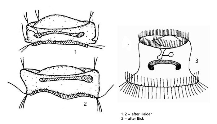
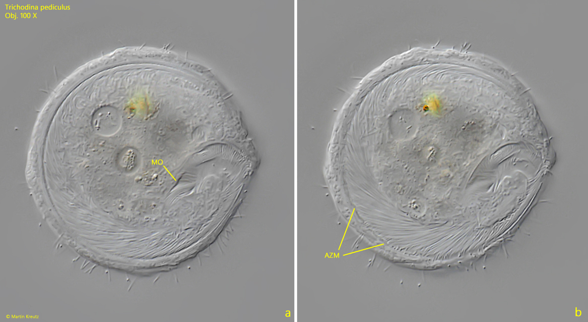
peristomial disc with ring-shaped adoral membranelle
cytostome at end of adoral membranelle
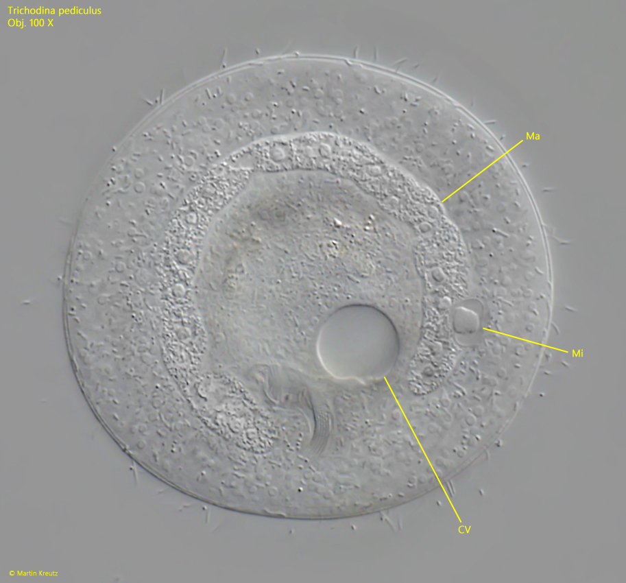
contractile vacuole near mid-body
macronucleus almost ring-shaped
one spherical micronucleus, adjacent to macronucleus
pellicle smooth, without rows of cilia
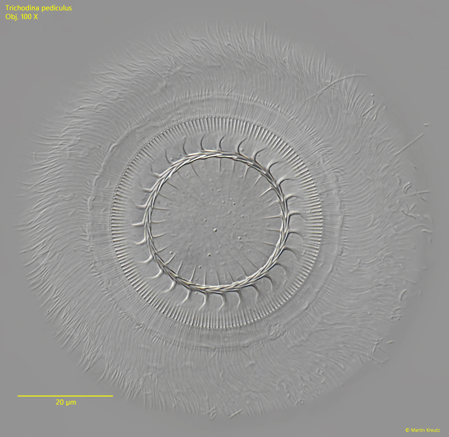
dorsal side with a complicate adhesive organelle
adhesive organelle consists of 3 ciliary wreaths and an adhesive disc
adhesive disc concave with denticle ring and radial pins
Trichodina pediculus
I mainly find Trichodina pediculus on Hydra viridis, because this is also the most common Hydra species in Simmelried. Only very occasionally have I found Trichodina pediculus on small water snails of unknown species. However, it is known that Trichodina pediculus also occurs on bryozoans, juvenile and adult fishes, and amphibian larvae.
Trichodina pediculus used to be classified as an ectoparasite because it was assumed that the ciliate could damage the fish skin with its adhesive organelle. However, the ciliate is now classified as an epibiont (Foissner, 1999). In fishes that are sick or stressed due to other causes, however, Trichodina pediculus can develop en masse, leading to skin damage and bacterial infection as a secondary reaction.
Hydra viridis can be colonized by several hundred Trichodina pediculus specimens without any negative effects on the host being observed. Trichodina pediculus moves very quickly across the host’s epidermis without losing its adhesion. Only when the Hydra viridis is slightly squeezed under the coverslip does Trichodina pediculus leave the host.
One might get the impression that Trichodina pediculus “grazes” on the host and is oriented toward the host with its mouth opening. However, this is not the case, because the adhesive organ is located on the dorsal side of the ciliate. This means that Trichodina pediculus glides over the host with its back. The mouth opening with the circular adoral zone is located on the side facing away from the host (s. figs. 3 and 4 a-b).
The composition of Trichodina pediculus‘s diet is largely unknown. I myself was able to identify mainly bacteria and small algae in the food vacuoles of specimens that lived on Hydra viridis. I did not observe any cells being detached from the epidermis of Hydra. I therefore consider it likely that Trichodina pediculus lives off the excreted food residues of the host.
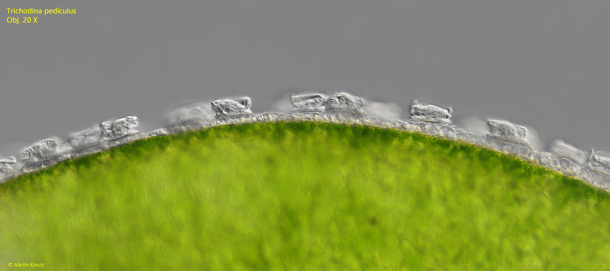
Fig. 1: Trichodina pediculus. D = 52–55 µm. Hundrets of specimens crawling over the body of Hydra viridis. Obj. 20 X.
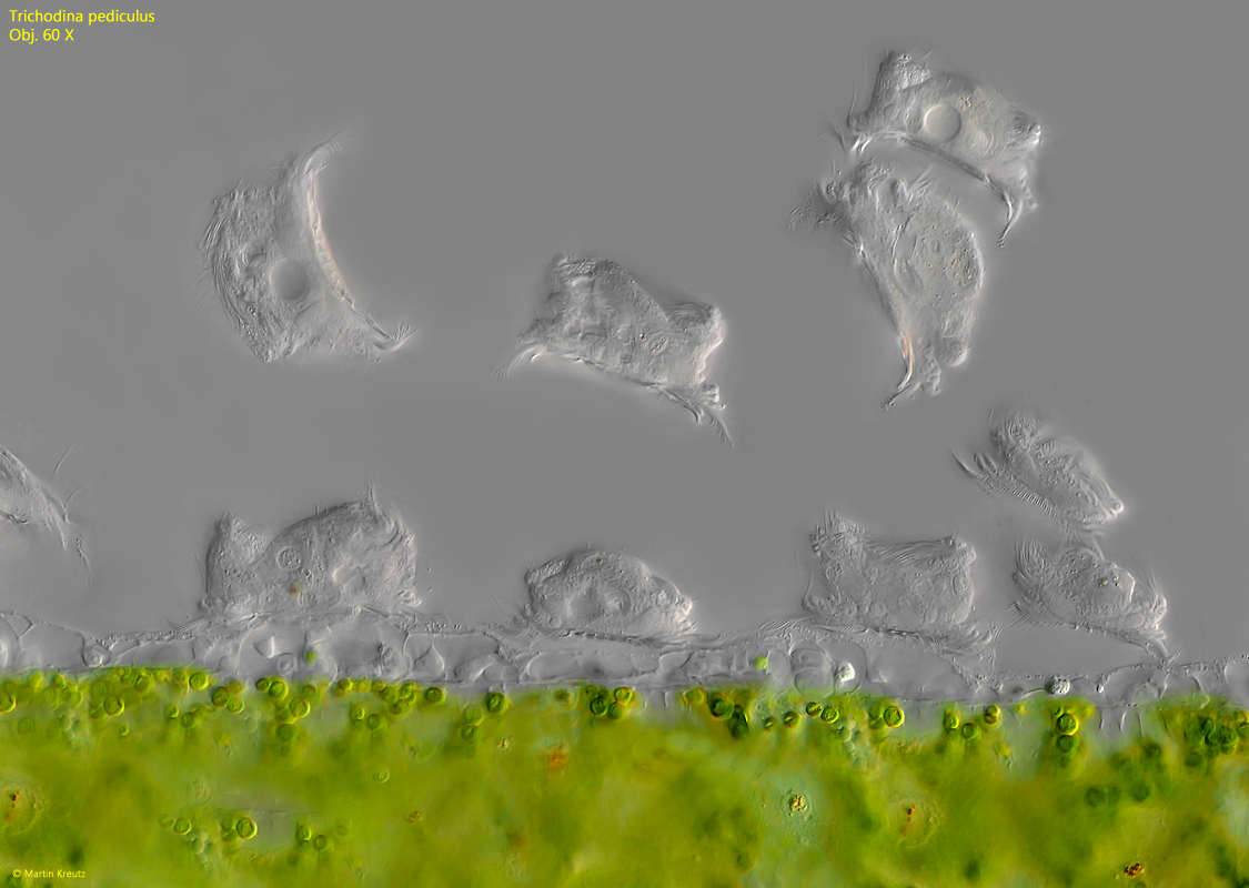
Fig. 2: Trichodina pediculus. D = 48–58 µm. The epidermis of Hydra viridis with some crawling and some detached specimens. Obj. 60 X.
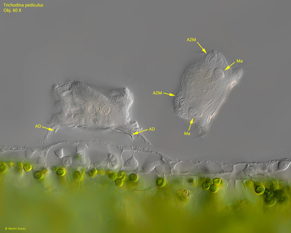
Fig. 3: Trichodina pediculus. D = 54–58 µm. A crawling and a detached specimen in lateral view. The crawling specimen is directed with the dorsal side to the epidermis of Hydra viridis, where the adhesive disc (AD) is located. In the detached specimen the adoral zone of membranelles (AZM) is visible on the ventral side and the ring-shaped macronucleus (Ma) is visible. Obj. 60 X.
Fig. 4 a-b: Trichodina pediculus. D = 48 µm. Two focal planes of a specimen from ventral. The ring-shaped adoral zone (AZM) ends in the mouth opening (MO). Obj. 100 X.
Fig. 5: Trichodina pediculus. A slightly squashed specimen from ventral with focal plane on the almost circular shaped macronucleus (Ma) with the adjacent micronucleus (Mi). CV = vontractile vacuole. Obj. 100 X.
Fig. 6: Trichodina pediculus. Dorsal view of a slightly squashed specimen with focal planes on the complicate shaped disc of the adhesive organelle. Obj. 100 X.