Although Trimyema compressum is described as a common species, I have only found a few specimens in Purren pond among decomposing leaves.
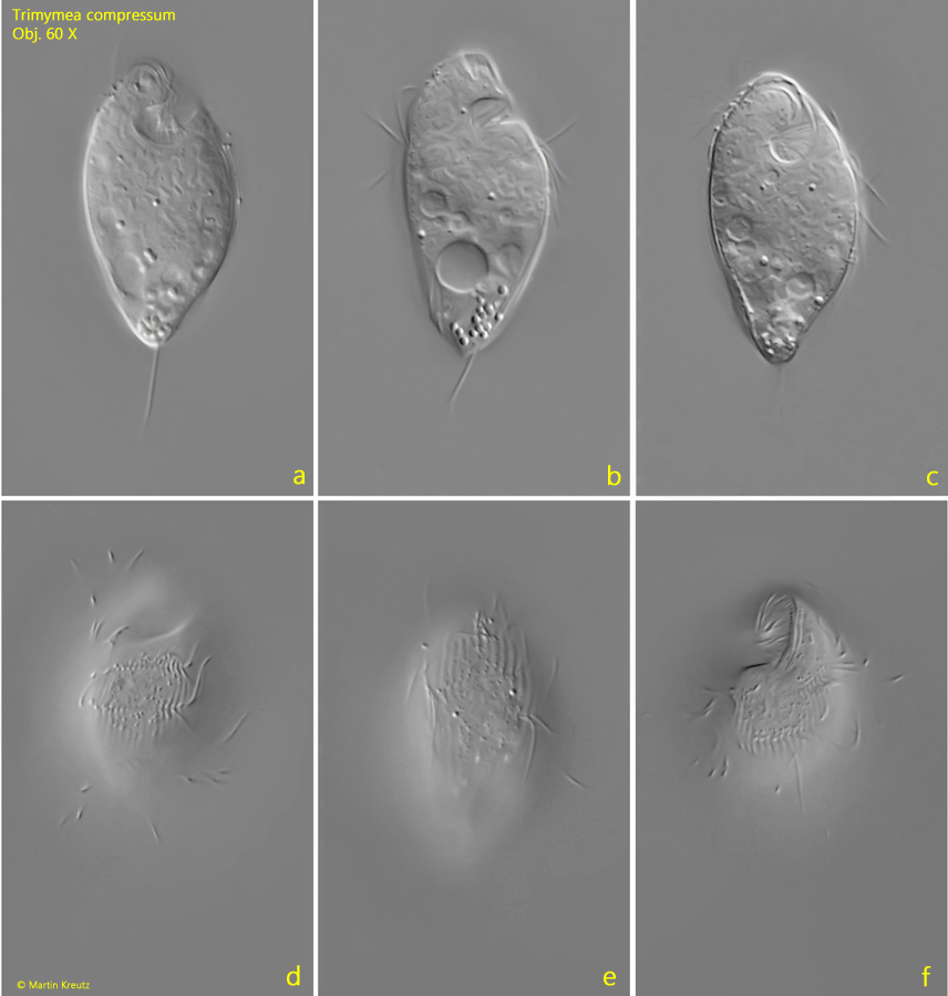
At low magnifications, the spindle-shaped ciliate appears obliquely truncated at the apical end. Here is the oral apparatus, which, from a ventral view, has the shape of a mirrored “6” (s. fig. 1 c). The anterior two-thirds of the body are covered with 50–60 longitudinal rows of cilia, which are divided into three transverse and slightly spiral bands. Between the longitudinal rows of cilia are longitudinal ribs, which are also divided into 3 bands (s. figs. 2 c and 3 a). This results in the typical ribbed structure of Trimyema compressum. The posterior third of the ciliate is naked, except for the caudal cilium.
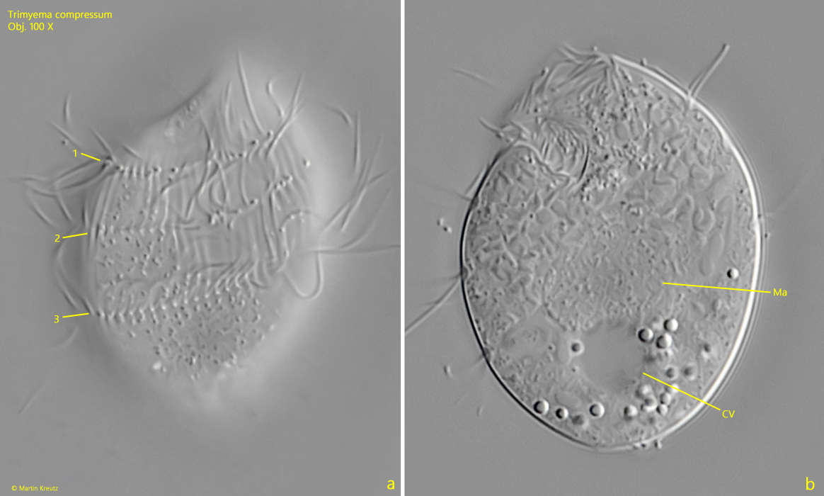
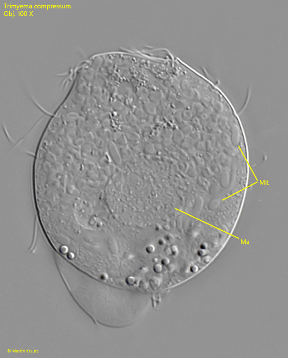
The macronucleus is round and was difficult to see in the living specimen (s. figs. 3 b and 4). I could not detect the micronucleus at all. It may only be visible after staining. In the cytoplasm, there are elongated structures measuring 3–4 µm in length, which I believe to be mitochondria because they have an irregular shape (s. fig. 4). However, Augustin, Foissner, and Adam (1987) consider these structures to be (symbiotic?) bacteria.
Fig. 1 a-f:Trimyema compressum. L = 45 µm. A freely swimming specimen from right (a, b), ventral (c, d), dorsal (e) and from left (f). Obj. 60 X.
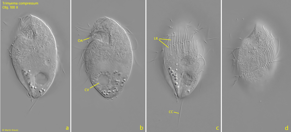
Fig. 1 a-f:Trimyema compressum. L = 45 µm. The slightly squashed specimen as shown in fig. 1 a-f from left. CC = caudal cilium, CV = contractile vacuole, OA = oral apparatus. Obj. 100 X.
Fig. 3 a-b:Trimyema compressum. A squashed specimen from left. Note the 3 spirally rows of cilia (1-3). CV = contractile vacuole, Ma = macronucleus. Obj. 100 X.
Fig. 4:Trimyema compressum. A strongly squashed specimen. Ma = macronucleus, Mit = mitochondria. Obj. 100 X.