macronucleus elliptical with one adjacent micronucleus
2–11 irregularly distributed contractile vacuoles
19–22 ventral somatic kineties
preoral kinety from mouth opening to the beak, not interrupted
2 circumoral kineties
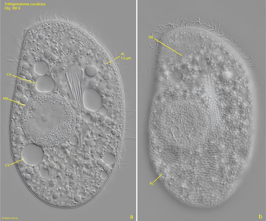
pellicles covered with alveolar layer, about 1 µm
alveolar layer with reticulate pattern
Trithigmostoma cucullulus
Trithigmostoma cucullulus is a very common ciliate that I find in almost all my localities. It is easy to observe as it likes to settle on the floating coverslip.
Trithigmostoma cucullulus has the typical structure of a cyrtophorid ciliate. The body is dorso-ventrally flattened. The dorsal side is weakly convex and naked with the exception of an anterior dorsal brush (s. figs. 1 b and 3). In contrast, the ventral side is flat and ciliated. In the anterior third the mouth opening with an oral basked is located. A pre-oral kinety extends from the mouth opening to the beak-shaped bulge of the body on the left side at the anterior end. The shape of this pre-oral kinety is an important identifying feature of cyrtophorid ciliates. In the genus Trithigmostoma it is continuous, without interruption. In addition, there is no cilium-free field in the middle of the ventral side, as is the case in the genus Chilodonella, for example. In Trithigmostoma cucullulus, the ventral somatic kinetes run approximately parallel and all end below or above the pre-oral kinety. This results in the typical cilia pattern of cyrtophorid ciliates (s. fig. 4).
Trithigmostoma cucullulus feeds mainly on diatoms, cyanobacteria and small algae, which are often gutted en masse. The cytoplasm contains 2–11 contractile vacuoles. In my population there were usually 5–6 (s. fig. 1 a). The macronucleus is ellipsoid with a central nucleolus. The micronucleus is close to the macronucleus (s. fig. 5).
The body of Trithigmostoma cucullulus is covered by an alveolar layer about 1 µm thick. This has a mucus-like character and has a very conspicuous, reticulate pattern (s. figs. 2 b and 6). This can be seen particularly well on the dorsal side. The alveolar layer can be shed and then often shows gaps.
Fig. 1 a-b:Trithigmostoma cucullulus. L = 128 µm. A freely swimming specimen from ventral (a) and from dorsal (b). Note the thin alveolar layer covering the body (AL) and the dorsal brush (DB). Ma = macronucleus, OB = oral basket. Obj. 100 X.
Fig. 2 a-b:Trithigmostoma cucullulus. L = 110 µm. Two focal planes of a second specimen from dorsal. Note the 1.3 µm thick alveolar layer (AL). Is the focal plane on the surface of the alveolar layer it shows a reticulate pattern (b). CV = contractile vacuoles, DB = dorsal brush, Ma = macronucleus. Obj. 100 X.
Fig. 3:Trithigmostoma cucullulus. A squashed specimen from right. While the dorsal side is naked apart from the dorsal brush the somatic kineties of the ventral side (VK) are visible. Obj. 100 X.
Fig. 4:Trithigmostoma cucullulus. L = 125 µm. The ventral side of a slightly squashed specimen. The somatic kineties (SOK) running parallel to the pre-oral kinety (POK). The oral pasked is surrounded by two circumoral kineties (COK). Obj. 100 X.
Fig. 5:Trithigmostoma cucullulus. The macronucleus (Ma) and the micronucleus (Mi) in a squashed specimen. Obj. 100 X.
Fig. 6:Trithigmostoma cucullulus. The reticulate pattern of the alveolar layer (RA) on the dorsal side of a squashed specimen. DB = dorsal brush. Obj. 100 X.