terminal cells broadly rounded, with slightly thickened membrane
Tychonema bornetii
I regularly find Tychonema bornetii in various locations. Most of the time, the cell filaments lie on the uppermost layer of mud or colonize decaying fallen leaves.
Tychonema bornetii was first described by Zukal in 1894 as Oscillatoria bornetii and was transferred to the genus Tychonema by Anagnostidis & Komárek in 1988. The main characteristic of Tychonema bornetii is the apparent absence of chlorophyll, which makes the cells appear almost colorless. In thick layers, however, mats of trichomes usually appear brownish.
The cells have an almost square shape and are strongly vacuolated. In my population, the cells were yellowish-brown in color (s. fig. 2). Although no green coloration from chlorophyll is visible, Tychonema bornetii is capable of photosynthesis. This is made possible by so-called phycobilisomes. These are protein complexes that contain chlorophyll a, phycocyanin, and phycoerythrin. This protein complex also utilizes the yellow and green portions of light and is extremely effective. Tychonema bornetii can therefore also grow in low-light environments.
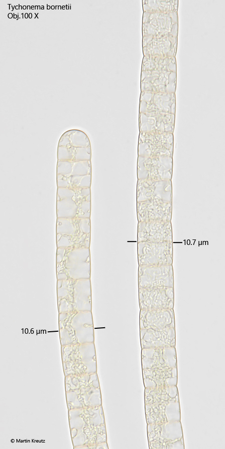
Contrary to the description by Huber-Pestalozzi (1938), I was also able to observe slight constrictions at the cross walls in my population (s. fig. 2). However, this was not visible in all trichomes. Possibly, this could also have been an effect of the cover slip pressure.
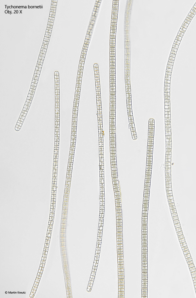
Fig. 1:Tychonema bornetii. Overview of several trichomes. Obj. 20 X.
Fig. 2:Tychonema bornetii. Two trichomes with a diameter of 10.6 µm and 10.7 µm in brightfield illumination. The terminal cell is broadly rounded. The cells are free from chlorophyll and have a yellowish-brownish color. At the crosswalls a slight constriction is visible. Obj. 100 X.
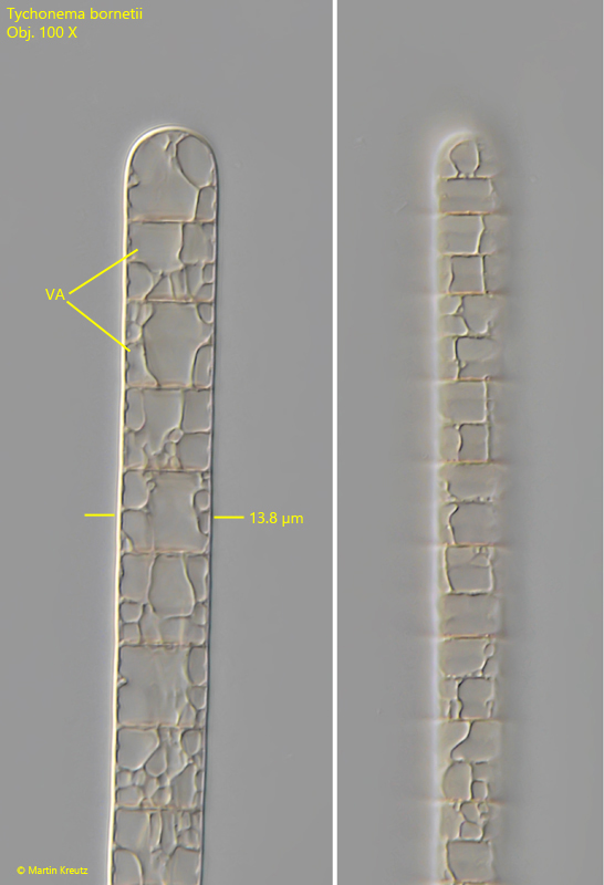
Fig. 3 a-b:Tychonema bornetii. Two focal planes of the end of a trichome with a diameter of 13.8 µm. The cells are strongly vacuolized (VA). Obj. 100 X.